FunctionPDZ and LIM domain proteins (PDLIM) contain PDZ and LIM domains. PDLIM binds α-actinin via its PDZ domain and other proteins via its LIM domain. PDLIM is implicated in cardiac and skeletal muscle structure[1].
For details on PDLIM see Group:MUZIC:ZASP and Group:MUZIC:Enigma Family.
- PDLIM1 promotes cell attachment at the cytoskeleton. For details see Group:MUZIC:ALP.
- PDLIM2 may have a role in bringing proteins to the cytoskeleton.
- PDLIM3 may have a role in organizing actin filament arrays within muscle cells.
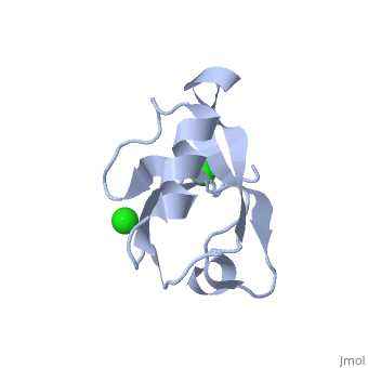
- PDLIM4 is a Zn+2 ion binding protein.
- PDLIM5 has important role in heart development by scaffolding PKC to the Z-disk region.
- PDLIM7 is an adaptor protein localizing the LIM-binding proteins to muscle tissues.
RelevancePDLIM2 may contribute to cancer cells migratory capacity. PDLIM5 overexpression promotes the development of heart hypertrophy.
| |
3D structures of PDZ and LIM domain protein3D structures of PDZ and LIM domain protein
Updated on 21-August-2023
{"openlevels":0}
- PDLIM1
- 2pkt – hPDLIM + α-actinin C terminal - human
- 1x62 – hPDLIM LIM domain - NMR
- PDLIM2
- 1vb7 – mPDLIM PDZ domain – mouse - NMR
- 2pa1 – hPDLIM PDZ domain + β-tropomysin C terminal
- 3pdv – hPDLIM PDZ domain + influenza virus NS1 C terminal
- PDLIM3
- 1v5l – mPDLIM PDZ domain - NMR
- 1x64 – mPDLIM LIM domain - NMR
- 4ydp - hPDLIM PDZ domain
- PDLIM4
- 4q2o – hPDLIM
- 2v1w – hPDLIM + α-actinin C terminal
- 2eeg – hPDLIM PDZ domain - NMR
- PDLIM5
- 1wf7 – mEnigma homolog protein PDZ domain - NMR
- 2dar – hEnigma homolog protein LIM domain - NMR
- 2uzc – hPDLIM + α-actinin C terminal
- PDLIM7
- 2q3g – hPDLIM PDZ domain + β-tropomysin C terminal
- 7rm8 – hPDLIM PDZ domain + SNX17 peptide - NMR
- PDLIM
- 1rgw – hPDLIM PDZ domain - NMR
ReferencesReferences
- ↑ te Velthuis AJ, Bagowski CP. PDZ and LIM domain-encoding genes: molecular interactions and their role in development. ScientificWorldJournal. 2007 Sep 1;7:1470-92. PMID:17767364 doi:10.1100/tsw.2007.232