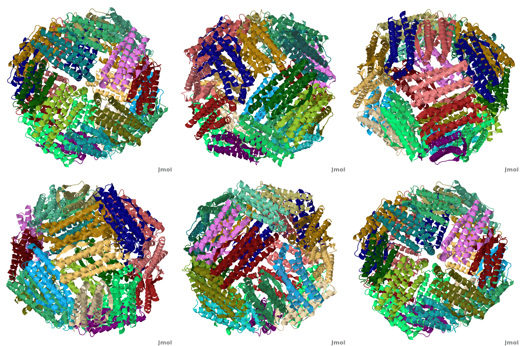
Ferritin
|
FunctionFunction
Ferritin (FR) is an iron storage and release protein. It stores iron as microcrystals with phosphate and hydroxide ions. FR is composed of 24 subunits of heavy chain (FTH) and light chain (FTL).[1] Amphibians have an additional middle subunit FR (FTM).
- Bacterioferritin (BFR) structure is very similar to FR. It contains a binuclear iron center and haem. It stores iron as ferric oxide mineral in its hollow central cavity.
- Thioferritin (TFR) is a ferritin-related protein with 2 cysteine residues adjacent to the dimetal binding site.
- Another iron storing protein is the DNA-binding Protein of Starved cells (Dps) - a ferritin-like diiron carboxylate.
- MrgA – another iron storage protein - belongs to the Dps family.
RelevanceRelevance
Cavities formed by FR are used for the manufacture of nanoparticles. FR is used as a marker for iron overload disorder.
DiseaseDisease
FR deficiency can lead to anemia.
3D Structures of Ferritin3D Structures of Ferritin
Updated on 23-August-2017
ReferencesReferences
- ↑ Wang W, Knovich MA, Coffman LG, Torti FM, Torti SV. Serum ferritin: Past, present and future. Biochim Biophys Acta. 2010 Aug;1800(8):760-9. doi: 10.1016/j.bbagen.2010.03.011. , Epub 2010 Mar 19. PMID:20304033 doi:http://dx.doi.org/10.1016/j.bbagen.2010.03.011
