1vpr
Crystal structure of a luciferase domain from the dinoflagellate Lingulodinium polyedrumCrystal structure of a luciferase domain from the dinoflagellate Lingulodinium polyedrum
Structural highlights
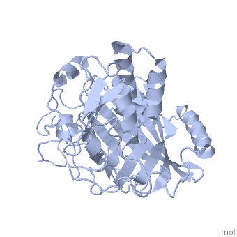
FunctionLUCIF_LINPO Emits blue light flashes with a wavelength of 475 nm during the night phase.[1] [2] [3] Evolutionary Conservation Check, as determined by ConSurfDB. You may read the explanation of the method and the full data available from ConSurf. Publication Abstract from PubMedThe luciferase of Lingulodinium polyedrum, a marine bioluminescent dinoflagellate, consists of three similar but not identical domains in a single polypeptide. Each encodes an active luciferase that catalyzes the oxidation of a chlorophyll-derived open tetrapyrrole (dinoflagellate luciferin) to produce blue light. These domains share no sequence similarity with any other in the GenBank database and no structural or motif similarity with any other luciferase. We report here the 1.8-A crystal structure of the third domain, D3, at pH 8, and a mechanism for its activity regulation by pH. D3 consists of two major structural elements: a beta-barrel pocket putatively for substrate binding and catalysis and a regulatory three-helix bundle. N-terminal histidine residues previously shown to regulate activity by pH are at the interface of the helices in the bundle. Molecular dynamics calculations indicate that, in response to changes in pH, these histidines could trigger a large molecular motion of the bundle, thereby exposing the active site to the substrate. Crystal structure of a pH-regulated luciferase catalyzing the bioluminescent oxidation of an open tetrapyrrole.,Schultz LW, Liu L, Cegielski M, Hastings JW Proc Natl Acad Sci U S A. 2005 Feb 1;102(5):1378-83. Epub 2005 Jan 21. PMID:15665092[4] From MEDLINE®/PubMed®, a database of the U.S. National Library of Medicine. See AlsoReferences
|
| ||||||||||||||||||