2f4m
The Mouse PNGase-HR23 Complex Reveals a Complete Remodulation of the Protein-Protein Interface Compared to its Yeast OrthologsThe Mouse PNGase-HR23 Complex Reveals a Complete Remodulation of the Protein-Protein Interface Compared to its Yeast Orthologs
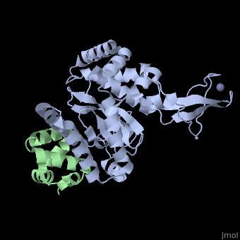
Structural highlights
Function[NGLY1_MOUSE] Specifically deglycosylates the denatured form of N-linked glycoproteins in the cytoplasm and assists their proteasome-mediated degradation. Cleaves the beta-aspartyl-glucosamine (GlcNAc) of the glycan and the amide side chain of Asn, converting Asn to Asp. Prefers proteins containing high-mannose over those bearing complex type oligosaccharides. Can recognize misfolded proteins in the endoplasmic reticulum that are exported to the cytosol to be destroyed and deglycosylate them, while it has no activity toward native proteins. Deglycosylation is a prerequisite for subsequent proteasome-mediated degradation of some, but not all, misfolded glycoproteins.[1] [2] [3] [RD23B_MOUSE] Multiubiquitin chain receptor involved in modulation of proteasomal degradation. Binds to polyubiquitin chains. Proposed to be capable to bind simultaneously to the 26S proteasome and to polyubiquitinated substrates and to deliver ubiquitinated proteins to the proteasome. May play a role in endoplasmic reticulum-associated degradation (ERAD) of misfolded glycoproteins by association with PNGase and delivering deglycosylated proteins to the proteasome.[4] [5] [6] Involved in global genome nucleotide excision repair (GG-NER) by acting as component of the XPC complex. Cooperatively with Cetn2 appears to stabilize Xpc. May protect Xpc from proteasomal degradation (By similarity).[7] [8] [9] The XPC complex is proposed to represent the first factor bound at the sites of DNA damage and together with other core recognition factors, Xpa, RPA and the TFIIH complex, is part of the pre-incision (or initial recognition) complex. The XPC complex recognizes a wide spectrum of damaged DNA characterized by distortions of the DNA helix such as single-stranded loops, mismatched bubbles or single stranded overhangs. The orientation of XPC complex binding appears to be crucial for inducing a productive NER. XPC complex is proposed to recognize and to interact with unpaired bases on the undamaged DNA strand which is followed by recruitment of the TFIIH complex and subsequent scanning for lesions in the opposite strand in a 5'-to-3' direction by the NER machinery. Cyclobutane pyrimidine dimers (CPDs) which are formed upon UV-induced DNA damage esacpe detection by the XPC complex due to a low degree of structural perurbation. Instead they are detected by the UV-DDB complex which in turn recruits and cooperates with the XPC complex in the respective DNA repair. In vitro, the Xpc:Rad23b dimer is sufficient to initiate NER; it preferentially binds to cisplatin and UV-damaged double-stranded DNA and also binds to a variety of chemically and structurally diverse DNA adducts. Xpc:Rad23b contacts DNA both 5' and 3' of a cisplatin lesion with a preference for the 5' side. Xpc:Rad23bB induces a bend in DNA upon binding. Xpc:Rad23b stimulates the activity of DNA glycosylases Tdg and Smug1 (By similarity).[10] [11] [12] Evolutionary Conservation Check, as determined by ConSurfDB. You may read the explanation of the method and the full data available from ConSurf. Publication Abstract from PubMedPeptide N-glycanase removes N-linked oligosaccharides from misfolded glycoproteins as part of the endoplasmic reticulum-associated degradation pathway. This process involves the formation of a tight complex of peptide N-glycanase with Rad23 in yeast and the orthologous HR23 proteins in mammals. In addition to its function in endoplasmic reticulum-associated degradation, HR23 is also involved in DNA repair, where it plays an important role in damage recognition in complex with the xeroderma pigmentosum group C protein. To characterize the dual role of HR23, we have determined the high resolution crystal structure of the mouse peptide N-glycanase catalytic core in complex with the xeroderma pigmentosum group C binding domain from HR23B. Peptide N-glycanase features a large cleft between its catalytic cysteine protease core and zinc binding domain. Opposite the zinc binding domain is the HR23B-interacting region, and surprisingly, the complex interface is fundamentally different from the orthologous yeast peptide N-glycanase-Rad23 complex. Different regions on both proteins are involved in complex formation, revealing an amazing degree of divergence in the interaction between two highly homologous proteins. Furthermore, the mouse peptide N-glycanase-HR23B complex mimics the interaction between xeroderma pigmentosum group C and HR23B, thereby providing a first structural model of how the two proteins interact within the nucleotide excision repair cascade in higher eukaryotes. The different interaction interfaces of the xeroderma pigmentosum group C binding domains in yeast and mammals suggest a co-evolution of the endoplasmic reticulum-associated degradation and DNA repair pathways. Structure of the mouse peptide N-glycanase-HR23 complex suggests co-evolution of the endoplasmic reticulum-associated degradation and DNA repair pathways.,Zhao G, Zhou X, Wang L, Li G, Kisker C, Lennarz WJ, Schindelin H J Biol Chem. 2006 May 12;281(19):13751-61. Epub 2006 Feb 24. PMID:16500903[13] From MEDLINE®/PubMed®, a database of the U.S. National Library of Medicine. See AlsoReferences
|
| ||||||||||||||||||||||