2emt: Difference between revisions
New page: left|200px {{Structure |PDB= 2emt |SIZE=350|CAPTION= <scene name='initialview01'>2emt</scene>, resolution 2.80Å |SITE= |LIGAND= |ACTIVITY= |GENE= }} '''Crys... |
No edit summary |
||
| Line 7: | Line 7: | ||
|ACTIVITY= | |ACTIVITY= | ||
|GENE= | |GENE= | ||
|DOMAIN= | |||
|RELATEDENTRY=[[1j19|1J19]], [[1gc6|1GC6]], [[1gc7|1GC7]], [[2d11|2D11]], [[2d10|2D10]], [[2d2q|2D2Q]], [[2ems|2EMS]] | |||
|RESOURCES=<span class='plainlinks'>[http://oca.weizmann.ac.il/oca-docs/fgij/fg.htm?mol=2emt FirstGlance], [http://oca.weizmann.ac.il/oca-bin/ocaids?id=2emt OCA], [http://www.ebi.ac.uk/pdbsum/2emt PDBsum], [http://www.rcsb.org/pdb/explore.do?structureId=2emt RCSB]</span> | |||
}} | }} | ||
| Line 30: | Line 33: | ||
[[Category: protein-peptide complex]] | [[Category: protein-peptide complex]] | ||
''Page seeded by [http://oca.weizmann.ac.il/oca OCA ] on | ''Page seeded by [http://oca.weizmann.ac.il/oca OCA ] on Mon Mar 31 02:51:13 2008'' | ||
Revision as of 02:51, 31 March 2008
| |||||||
| , resolution 2.80Å | |||||||
|---|---|---|---|---|---|---|---|
| Related: | 1J19, 1GC6, 1GC7, 2D11, 2D10, 2D2Q, 2EMS
| ||||||
| Resources: | FirstGlance, OCA, PDBsum, RCSB | ||||||
| Coordinates: | save as pdb, mmCIF, xml | ||||||
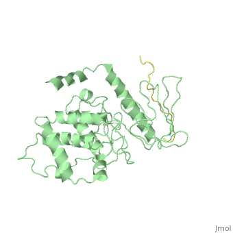
Crystal Structure Analysis of the radixin FERM domain complexed with adhesion molecule PSGL-1
OverviewOverview
P-selectin glycoprotein ligand-1 (PSGL-1), an adhesion molecule with O-glycosylated extracellular sialomucins, is involved in leukocyte inflammatory responses. On activation, ezrin-radixin-moesin (ERM) proteins mediate the redistribution of PSGL-1 on polarized cell surfaces to facilitate binding to target molecules. ERM proteins recognize a short binding motif, Motif-1, conserved in cytoplasmic tails of adhesion molecules, whereas PSGL-1 lacks Motif-1 residues important for binding to ERM proteins. The crystal structure of the complex between the radixin FERM domain and a PSGL-1 juxtamembrane peptide reveals that the peptide binds the groove of FERM subdomain C by forming a beta-strand associated with strand beta5C, followed by a loop flipped out towards the solvent. The Motif-1 3(10) helix present in the FERM-ICAM-2 complex is absent in PSGL-1 given the absence of a critical Motif-1 alanine residue, and PSGL-1 reduces its contact area with subdomain C. Non-conserved positions are occupied by large residues Met9 and His8, which stabilize peptide conformation and enhance groove binding. Non-conserved residues play an important role in compensating for loss of binding energy resulting from the absence of conserved residues important for binding.
About this StructureAbout this Structure
2EMT is a Protein complex structure of sequences from Mus musculus. Full crystallographic information is available from OCA.
ReferenceReference
Structural basis of PSGL-1 binding to ERM proteins., Takai Y, Kitano K, Terawaki S, Maesaki R, Hakoshima T, Genes Cells. 2007 Dec;12(12):1329-38. PMID:18076570
Page seeded by OCA on Mon Mar 31 02:51:13 2008