4op0: Difference between revisions
No edit summary |
No edit summary |
||
| Line 3: | Line 3: | ||
== Structural highlights == | == Structural highlights == | ||
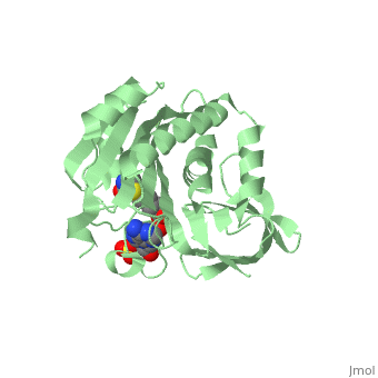
<table><tr><td colspan='2'>[[4op0]] is a 2 chain structure with sequence from [http://en.wikipedia.org/wiki/Myctu Myctu]. Full crystallographic information is available from [http://oca.weizmann.ac.il/oca-bin/ocashort?id=4OP0 OCA]. For a <b>guided tour on the structure components</b> use [http://oca.weizmann.ac.il/oca-docs/fgij/fg.htm?mol=4OP0 FirstGlance]. <br> | <table><tr><td colspan='2'>[[4op0]] is a 2 chain structure with sequence from [http://en.wikipedia.org/wiki/Myctu Myctu]. Full crystallographic information is available from [http://oca.weizmann.ac.il/oca-bin/ocashort?id=4OP0 OCA]. For a <b>guided tour on the structure components</b> use [http://oca.weizmann.ac.il/oca-docs/fgij/fg.htm?mol=4OP0 FirstGlance]. <br> | ||
</td></tr><tr><td class="sblockLbl"><b>[[Ligand|Ligands:]]</b></td><td class="sblockDat"><scene name='pdbligand=BT5:BIOTINYL-5-AMP'>BT5</scene>, <scene name='pdbligand=SO4:SULFATE+ION'>SO4</scene>< | </td></tr><tr id='ligand'><td class="sblockLbl"><b>[[Ligand|Ligands:]]</b></td><td class="sblockDat"><scene name='pdbligand=BT5:BIOTINYL-5-AMP'>BT5</scene>, <scene name='pdbligand=SO4:SULFATE+ION'>SO4</scene></td></tr> | ||
<tr><td class="sblockLbl"><b>[[Gene|Gene:]]</b></td><td class="sblockDat">birA, MT3379, Rv3279c ([http://www.ncbi.nlm.nih.gov/Taxonomy/Browser/wwwtax.cgi?mode=Info&srchmode=5&id=83332 MYCTU])</td></tr> | <tr id='gene'><td class="sblockLbl"><b>[[Gene|Gene:]]</b></td><td class="sblockDat">birA, MT3379, Rv3279c ([http://www.ncbi.nlm.nih.gov/Taxonomy/Browser/wwwtax.cgi?mode=Info&srchmode=5&id=83332 MYCTU])</td></tr> | ||
<tr><td class="sblockLbl"><b>Activity:</b></td><td class="sblockDat"><span class='plainlinks'>[http://en.wikipedia.org/wiki/Biotin--[acetyl-CoA-carboxylase]_ligase Biotin--[acetyl-CoA-carboxylase] ligase], with EC number [http://www.brenda-enzymes.info/php/result_flat.php4?ecno=6.3.4.15 6.3.4.15] </span></td></tr> | <tr id='activity'><td class="sblockLbl"><b>Activity:</b></td><td class="sblockDat"><span class='plainlinks'>[http://en.wikipedia.org/wiki/Biotin--[acetyl-CoA-carboxylase]_ligase Biotin--[acetyl-CoA-carboxylase] ligase], with EC number [http://www.brenda-enzymes.info/php/result_flat.php4?ecno=6.3.4.15 6.3.4.15] </span></td></tr> | ||
<tr><td class="sblockLbl"><b>Resources:</b></td><td class="sblockDat"><span class='plainlinks'>[http://oca.weizmann.ac.il/oca-docs/fgij/fg.htm?mol=4op0 FirstGlance], [http://oca.weizmann.ac.il/oca-bin/ocaids?id=4op0 OCA], [http://www.rcsb.org/pdb/explore.do?structureId=4op0 RCSB], [http://www.ebi.ac.uk/pdbsum/4op0 PDBsum]</span></td></tr> | <tr id='resources'><td class="sblockLbl"><b>Resources:</b></td><td class="sblockDat"><span class='plainlinks'>[http://oca.weizmann.ac.il/oca-docs/fgij/fg.htm?mol=4op0 FirstGlance], [http://oca.weizmann.ac.il/oca-bin/ocaids?id=4op0 OCA], [http://www.rcsb.org/pdb/explore.do?structureId=4op0 RCSB], [http://www.ebi.ac.uk/pdbsum/4op0 PDBsum]</span></td></tr> | ||
<table> | </table> | ||
<div style="background-color:#fffaf0;"> | <div style="background-color:#fffaf0;"> | ||
== Publication Abstract from PubMed == | == Publication Abstract from PubMed == | ||
| Line 21: | Line 21: | ||
</StructureSection> | </StructureSection> | ||
[[Category: Myctu]] | [[Category: Myctu]] | ||
[[Category: Akhter, Y | [[Category: Akhter, Y]] | ||
[[Category: Ma, Q | [[Category: Ma, Q]] | ||
[[Category: Wilmanns, M | [[Category: Wilmanns, M]] | ||
[[Category: Bira]] | [[Category: Bira]] | ||
[[Category: Ligase]] | [[Category: Ligase]] | ||
Revision as of 12:15, 5 January 2015
Crystal structure of biotin protein ligase (RV3279C) of Mycobacterium tuberculosis, complexed with biotinyl-5'-AMPCrystal structure of biotin protein ligase (RV3279C) of Mycobacterium tuberculosis, complexed with biotinyl-5'-AMP
Structural highlights
Publication Abstract from PubMedProtein biotinylation, a rare form of post-translational modification, is found in enzymes required for lipid biosynthesis. In mycobacteria, this process is essential for the formation of their complex and distinct cell wall and has become a focal point of drug discovery approaches. The enzyme responsible for this process, biotin protein ligase, substantially varies in different species in terms of overall structural organization, regulation of function and substrate specificity. To advance the understanding of the molecular mechanism of biotinylation in Mycobacterium tuberculosis we have biochemically and structurally characterized the corresponding enzyme. We report the high-resolution crystal structures of the apo-form and reaction intermediate biotinyl-5'-AMP-bound form of M. tuberculosis biotin protein ligase. Binding of the reaction intermediate leads to clear disorder-to-order transitions. We show that a conserved lysine, Lys138, in the active site is essential for biotinylation. Active site conformational changes upon reaction intermediate biotinyl-5'-AMP binding in biotin protein ligase from Mycobacterium tuberculosis.,Ma Q, Akhter Y, Wilmanns M, Ehebauer MT Protein Sci. 2014 Apr 9. doi: 10.1002/pro.2475. PMID:24723382[1] From MEDLINE®/PubMed®, a database of the U.S. National Library of Medicine. References
|
| ||||||||||||||||||||