2emt: Difference between revisions
No edit summary |
No edit summary |
||
| Line 3: | Line 3: | ||
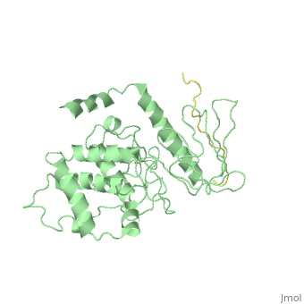
<StructureSection load='2emt' size='340' side='right'caption='[[2emt]], [[Resolution|resolution]] 2.80Å' scene=''> | <StructureSection load='2emt' size='340' side='right'caption='[[2emt]], [[Resolution|resolution]] 2.80Å' scene=''> | ||
== Structural highlights == | == Structural highlights == | ||
<table><tr><td colspan='2'>[[2emt]] is a 5 chain structure with sequence from [ | <table><tr><td colspan='2'>[[2emt]] is a 5 chain structure with sequence from [https://en.wikipedia.org/wiki/Lk3_transgenic_mice Lk3 transgenic mice]. Full crystallographic information is available from [http://oca.weizmann.ac.il/oca-bin/ocashort?id=2EMT OCA]. For a <b>guided tour on the structure components</b> use [https://proteopedia.org/fgij/fg.htm?mol=2EMT FirstGlance]. <br> | ||
</td></tr><tr id='related'><td class="sblockLbl"><b>[[Related_structure|Related:]]</b></td><td class="sblockDat">[[1j19|1j19]], [[1gc6|1gc6]], [[1gc7|1gc7]], [[2d11|2d11]], [[2d10|2d10]], [[2d2q|2d2q]], [[2ems|2ems]]</td></tr> | </td></tr><tr id='related'><td class="sblockLbl"><b>[[Related_structure|Related:]]</b></td><td class="sblockDat"><div style='overflow: auto; max-height: 3em;'>[[1j19|1j19]], [[1gc6|1gc6]], [[1gc7|1gc7]], [[2d11|2d11]], [[2d10|2d10]], [[2d2q|2d2q]], [[2ems|2ems]]</div></td></tr> | ||
<tr id='resources'><td class="sblockLbl"><b>Resources:</b></td><td class="sblockDat"><span class='plainlinks'>[ | <tr id='resources'><td class="sblockLbl"><b>Resources:</b></td><td class="sblockDat"><span class='plainlinks'>[https://proteopedia.org/fgij/fg.htm?mol=2emt FirstGlance], [http://oca.weizmann.ac.il/oca-bin/ocaids?id=2emt OCA], [https://pdbe.org/2emt PDBe], [https://www.rcsb.org/pdb/explore.do?structureId=2emt RCSB], [https://www.ebi.ac.uk/pdbsum/2emt PDBsum], [https://prosat.h-its.org/prosat/prosatexe?pdbcode=2emt ProSAT]</span></td></tr> | ||
</table> | </table> | ||
== Function == | == Function == | ||
[[ | [[https://www.uniprot.org/uniprot/RADI_MOUSE RADI_MOUSE]] Probably plays a crucial role in the binding of the barbed end of actin filaments to the plasma membrane. [[https://www.uniprot.org/uniprot/SELPL_MOUSE SELPL_MOUSE]] A SLe(x)-type glycan, which through high affinity, calcium-dependent interactions with E- and P-selectins, mediates rapid rolling of leukocytes over vascular surfaces during the initial steps in inflammation. PSGL1 is critical for the initial leukocyte capture.<ref>PMID:11104809</ref> <ref>PMID:12370362</ref> <ref>PMID:17442598</ref> | ||
== Evolutionary Conservation == | == Evolutionary Conservation == | ||
[[Image:Consurf_key_small.gif|200px|right]] | [[Image:Consurf_key_small.gif|200px|right]] | ||
Revision as of 12:50, 5 May 2021
Crystal Structure Analysis of the radixin FERM domain complexed with adhesion molecule PSGL-1Crystal Structure Analysis of the radixin FERM domain complexed with adhesion molecule PSGL-1
Structural highlights
Function[RADI_MOUSE] Probably plays a crucial role in the binding of the barbed end of actin filaments to the plasma membrane. [SELPL_MOUSE] A SLe(x)-type glycan, which through high affinity, calcium-dependent interactions with E- and P-selectins, mediates rapid rolling of leukocytes over vascular surfaces during the initial steps in inflammation. PSGL1 is critical for the initial leukocyte capture.[1] [2] [3] Evolutionary Conservation Check, as determined by ConSurfDB. You may read the explanation of the method and the full data available from ConSurf. Publication Abstract from PubMedP-selectin glycoprotein ligand-1 (PSGL-1), an adhesion molecule with O-glycosylated extracellular sialomucins, is involved in leukocyte inflammatory responses. On activation, ezrin-radixin-moesin (ERM) proteins mediate the redistribution of PSGL-1 on polarized cell surfaces to facilitate binding to target molecules. ERM proteins recognize a short binding motif, Motif-1, conserved in cytoplasmic tails of adhesion molecules, whereas PSGL-1 lacks Motif-1 residues important for binding to ERM proteins. The crystal structure of the complex between the radixin FERM domain and a PSGL-1 juxtamembrane peptide reveals that the peptide binds the groove of FERM subdomain C by forming a beta-strand associated with strand beta5C, followed by a loop flipped out towards the solvent. The Motif-1 3(10) helix present in the FERM-ICAM-2 complex is absent in PSGL-1 given the absence of a critical Motif-1 alanine residue, and PSGL-1 reduces its contact area with subdomain C. Non-conserved positions are occupied by large residues Met9 and His8, which stabilize peptide conformation and enhance groove binding. Non-conserved residues play an important role in compensating for loss of binding energy resulting from the absence of conserved residues important for binding. Structural basis of PSGL-1 binding to ERM proteins.,Takai Y, Kitano K, Terawaki S, Maesaki R, Hakoshima T Genes Cells. 2007 Dec;12(12):1329-38. PMID:18076570[4] From MEDLINE®/PubMed®, a database of the U.S. National Library of Medicine. See AlsoReferences
|
| ||||||||||||||||