2v7f: Difference between revisions
No edit summary |
No edit summary |
||
| Line 1: | Line 1: | ||
==Structure of P. abyssi RPS19 protein== | ==Structure of P. abyssi RPS19 protein== | ||
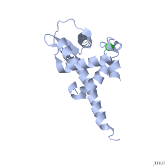
<StructureSection load='2v7f' size='340' side='right' caption='[[2v7f]], [[Resolution|resolution]] 1.15Å' scene=''> | <StructureSection load='2v7f' size='340' side='right'caption='[[2v7f]], [[Resolution|resolution]] 1.15Å' scene=''> | ||
== Structural highlights == | == Structural highlights == | ||
<table><tr><td colspan='2'>[[2v7f]] is a 1 chain structure with sequence from [http://en.wikipedia.org/wiki/"pyrococcus_abyssi"_erauso_et_al._1993 "pyrococcus abyssi" erauso et al. 1993]. Full crystallographic information is available from [http://oca.weizmann.ac.il/oca-bin/ocashort?id=2V7F OCA]. For a <b>guided tour on the structure components</b> use [http:// | <table><tr><td colspan='2'>[[2v7f]] is a 1 chain structure with sequence from [http://en.wikipedia.org/wiki/"pyrococcus_abyssi"_erauso_et_al._1993 "pyrococcus abyssi" erauso et al. 1993]. Full crystallographic information is available from [http://oca.weizmann.ac.il/oca-bin/ocashort?id=2V7F OCA]. For a <b>guided tour on the structure components</b> use [http://proteopedia.org/fgij/fg.htm?mol=2V7F FirstGlance]. <br> | ||
</td></tr><tr id='ligand'><td class="sblockLbl"><b>[[Ligand|Ligands:]]</b></td><td class="sblockDat"><scene name='pdbligand=CL:CHLORIDE+ION'>CL</scene></td></tr> | </td></tr><tr id='ligand'><td class="sblockLbl"><b>[[Ligand|Ligands:]]</b></td><td class="sblockDat" id="ligandDat"><scene name='pdbligand=CL:CHLORIDE+ION'>CL</scene></td></tr> | ||
<tr id='resources'><td class="sblockLbl"><b>Resources:</b></td><td class="sblockDat"><span class='plainlinks'>[http:// | <tr id='resources'><td class="sblockLbl"><b>Resources:</b></td><td class="sblockDat"><span class='plainlinks'>[http://proteopedia.org/fgij/fg.htm?mol=2v7f FirstGlance], [http://oca.weizmann.ac.il/oca-bin/ocaids?id=2v7f OCA], [http://pdbe.org/2v7f PDBe], [http://www.rcsb.org/pdb/explore.do?structureId=2v7f RCSB], [http://www.ebi.ac.uk/pdbsum/2v7f PDBsum], [http://prosat.h-its.org/prosat/prosatexe?pdbcode=2v7f ProSAT]</span></td></tr> | ||
</table> | </table> | ||
== Function == | == Function == | ||
| Line 36: | Line 36: | ||
</StructureSection> | </StructureSection> | ||
[[Category: Pyrococcus abyssi erauso et al. 1993]] | [[Category: Pyrococcus abyssi erauso et al. 1993]] | ||
[[Category: Large Structures]] | |||
[[Category: Aguissa-Toure, A H]] | [[Category: Aguissa-Toure, A H]] | ||
[[Category: Fribourg, S]] | [[Category: Fribourg, S]] | ||
Revision as of 14:22, 17 June 2020
Structure of P. abyssi RPS19 proteinStructure of P. abyssi RPS19 protein
Structural highlights
Function[RS19E_PYRAB] May be involved in maturation of the 30S ribosomal subunit. Evolutionary Conservation Check, as determined by ConSurfDB. You may read the explanation of the method and the full data available from ConSurf. Publication Abstract from PubMedDiamond-Blackfan anemia (DBA) is a rare congenital disease linked to mutations in the ribosomal protein genes rps19, rps24 and rps17. It belongs to the emerging class of ribosomal disorders. To understand the impact of DBA mutations on RPS19 function, we have solved the crystal structure of RPS19 from Pyrococcus abyssi. The protein forms a five alpha-helix bundle organized around a central amphipathic alpha-helix, which corresponds to the DBA mutation hot spot. From the structure, we classify DBA mutations relative to their respective impact on protein folding (class I) or on surface properties (class II). Class II mutations cluster into two conserved basic patches. In vivo analysis in yeast demonstrates an essential role for class II residues in the incorporation into pre-40S ribosomal particles. This data indicate that missense mutations in DBA primarily affect the capacity of the protein to be incorporated into pre-ribosomes, thus blocking maturation of the pre-40S particles. Molecular basis of Diamond-Blackfan anemia: structure and function analysis of RPS19.,Gregory LA, Aguissa-Toure AH, Pinaud N, Legrand P, Gleizes PE, Fribourg S Nucleic Acids Res. 2007;35(17):5913-21. Epub 2007 Aug 28. PMID:17726054[1] From MEDLINE®/PubMed®, a database of the U.S. National Library of Medicine. See AlsoReferences
|
| ||||||||||||||||