Nitric Oxide Synthase: Difference between revisions
Michal Harel (talk | contribs) No edit summary |
|||
| (205 intermediate revisions by 10 users not shown) | |||
| Line 1: | Line 1: | ||
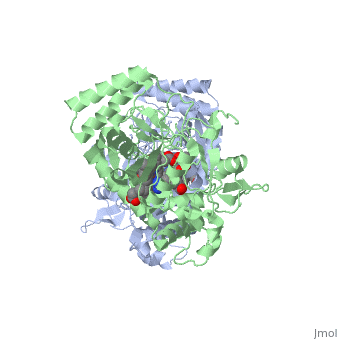
== | <StructureSection load='2g6h' size='350' side='right' caption='Neuronal nitric oxide synthase dimer complex with cofactor tetrahydrobiopterin, acetate and Zn+2 (grey), (PDB entry [[2g6h]])' scene=''> | ||
Nitric Oxide Synthase (NOS) is | '''Nitric Oxide Synthase''' (NOS) or '''Nitric Oxide Synthase oxygenase''' is an enzyme catalysing the formation of L-citrulline and [http://en.wikipedia.org/wiki/Nitric_Oxide/ nitric oxide] (NO) from L-arginine. NOS is a homodimeric protein with 125- to 160-kDa per monomer. In mammals, NOS appears as 3 isozymes: neuronal NOS (nNOS) (for details see [[Nos1]]), cytokine-inducible NOS (iNOS) and endothelial NOS (eNOS). The N-terminal domain of NOS is an '''oxygenase''' domain (OD). NOS cofactors are: NADPH, FAD, FMN, heme and O2. See also [[Nos1]]. | ||
=Introduction= | |||
[[Image: | In mammals three isozymes of NOS have been identified: Neuronal NOS (nNOS), inducible NOS (iNOS), and endothelial NOS (eNOS)<ref>Flemming, I., Chapter 3, Biology of Nitric Oxide Synthases, from Microcirculation, Editors Ronald F. Tuma, R. F., Durán, W. N., and Ley, K., 2nd edition, Academic Press, 2008</ref>. (The NOS enzymes are found in numeral organisms. Most facts used here are from the human NOS, but sites from different organisms are used). Neuronal NOS is producing NO in the nervous tissue in both the peripheral and the central nervous system. nNOS is functioning in cell signaling and communication - a vital part of the nervous tissue. Inducible NOS is connected with the immune system. Endothelial NOS is controlling the amount of NO signaling in the endothelial cells eg. blood vessel dilation. An overview of the structural organization of the NOS homodimer is given below. All cofactors are included and the electron transfer pathway which takes place in NOS is indicated. | ||
{{Clear}} | |||
[[Image:schematic_NOS.png|left|thumb|450px]] | |||
{{Clear}} | |||
Each NOS monomer is composed of two domains: an N-terminal oxygenase domain and a C-terminal reductase domain. Each subunit is held together by a zinc ion, which is bound by two cysteines from each oxygenase domain. Calmodulin binds to a linker region between the two domains and is necessary for activity. The reductase domain supplies electrons for the NOS reaction which takes place in the oxygenase domain. The reductase domain contains two redox-active prosthetic groups, flavin adenine dinucleotide (FAD) and Flavin mononucleotide (FMN). Nicotinamide adenine dinucleotide phosphate(NADPH) binds to the domain and passes on an electron to FAD which passes the electron on to FMN. FMN passes the electron on to the heme in the oxygenase domain on the opposite subunit. The oxygenase domain contains 5,6,7,8-tetrahydrobiopterin (H<sub>4</sub>B) and the already mentioned heme. These two are also redox active groups. Besides Heme and H<sub>4</sub>B, the oxygenase domain binds the substrate L-arginine which takes part in the NO synthase reaction (see below). | |||
<br/> | |||
Many structures of NOS have been determined. A list with additional information can be seen [[NOS structures | here]].<br/> | |||
== The reaction of NOS == | == The reaction of NOS == | ||
As previously described NOS is an enzyme | As previously described NOS is an enzyme consisting of two domains; the N-terminal oxygenase domain and the C-terminal reductase domain. | ||
The oxygenase domain | The NO production takes plase in the oxygenase domain, whereas the reductase domain provides the electrons necessary to drive the reaction in the oxygenase domain. | ||
The reaction is: | The reaction is: | ||
[[image:Makroprojekt-chemdraw-1-.gif|frame|left|thumb|400px]] | |||
{{Clear}} | |||
The reaction occurs in two steps. In the first step L-arginine is turned in to N-hydroxy-arginine. In the second step L-citrulline and NO are formed. The reaction utilizes 2O<sub>2</sub> and 3 electrons from 3/2 NADPH.<ref>PMID:17014963</ref> | |||
== The Oxygenase domain of NOS == | |||
[ | The <scene name='Sandbox_5/Nos_oxygenase_med_cofaktore/3'>oxygenase domain</scene> contains the active site of the enzyme. The active site binds the substrate <scene name='Nitric_oxide_synthase/Nos_oxygenase_arg/3'>L-Arginine</scene>([http://en.wikipedia.org/wiki/Arginine Arginine]) which is converted into citruline and NO (explained in details below). The domain has three cofactors bound: | ||
<scene name='35/351835/1/3'>Tetrahydrobiopterin</scene> ([http://en.wikipedia.org/wiki/BH4 (6R).5,6,7,8-Tetrahydrobiopterin]) | |||
<scene name='35/351835/1/4'>Heme</scene> ([http://en.wikipedia.org/wiki/Heme Heme]) | |||
= | <scene name='35/351835/Zinc/2'>Zinc ion</scene> | ||
The <scene name=' | === General Structure === | ||
The oxygenase domain can be divided into three subdomains. Firstly, the | |||
<scene name='Nitric_oxide_synthase/Oxygenase_subdomains/1'>substrate-binding subdomain</scene>, which is <font color='#3DFF00'>'''crescent in shape'''</font>, binds the substrate in an interior pocket of the crescent. The Heme group is located between the tips of the crescent shape, thus closing the pocket in which the substrate is located. Secondly the <scene name='Nitric_oxide_synthase/Oxygenase_subdomains/3'> H4B binding subdomain</scene> serves as a cap for the cavity created by the substrate binding domains crescent shape. Thirdly there is a subdomain consisting of <scene name='Nitric_oxide_synthase/Oxygenase_subdomains/2'>a two helix bundle</scene> making up a hydrophobic core, however this subdomain is not thought to take part in the catalytic activity of the enzyme. | |||
=== Substrate binding === | |||
<scene name=' | The active site is highly conserved in the different NOS species. Thus it is possible to discuss substrate binding i general terms. The NOS enzyme binds its substrate (L-arginine) in the distal pocket by hydrogen bindings both to the guanidino[http://en.wikipedia.org/wiki/Guanidino] end and the amino acid end. <scene name='Nitric_oxide_synthase/Substratebinding_test/1'>Substrate in the catalytic site</scene> is shown in green with the heme and H<sub>4</sub>B shown. NOS binds its substrate by coordinating CO(or O<sub>2</sub>) to the heme at the site occupied by oxygen<ref>PMID:9376373 </ref>(it is the opposite site of the Cys coordination to heme - look in the 'heme' section). The binding of substrate leads to a 2-step transformation first to N-hydroxy-L-arginine (the tightly bound intermediate) and then NO and L-Citrulline. The product NO can then either diffuse out of the <scene name='Nitric_oxide_synthase/Substratebinding_distal_pocket/1'>cavity</scene> or bind to the heme and function in NO auto-inhibition though this inhibition is diverse throughout the 3 isoforms<ref>PMID:15598509</ref>. | ||
PDB structures used in the section above: [[3nos]], [[2g6h]] | |||
---- | |||
==== | ===H<sub>4</sub>B=== | ||
The | [[image:bh4.png|left|frame|Structure of tetrahydrobiopterin]] | ||
{{Clear}} | |||
<scene name='Sandbox_5/Nos_oxygenase_bh4/11'>H4B</scene> is an essential cofactor in NOS and in the [[aromatic amino acid hydroxylases]]. NOS contains two molecules of <scene name='Sandbox_5/Begge_h4b/1'>H4B</scene>, one in each monomer. The active site forms part of the cavity already described. This cavity can be visualized as a <scene name='Nitric_oxide_synthase/Substratebinding_distal_pocket/2'>tunnel</scene>. Here H<sub>4</sub>B helps substrate interactions by lining the active-center tunnel and hydrogen bonding to the heme propionate and to alfa helix 7. The propionate group and alpha helix 7 are also involved in the L-Arg binding. This gives H<sub>4</sub>B the opportunity to play an important role in the control of subunit interactions and active site formation. H<sub>4</sub>B is therefore more or less a structural cofactor and has a stabilizing effect. Its structural importance is further reconned to play a role in dimer formation (dimerization requires bound zinc ion along with H<sub>4</sub>B), and major conformational changes leading to the formation of the active site channelform<ref name="Raman">PMID:9875848</ref>. | |||
[[image:H4b_hydrogenbindinger2.png|thumb|left|Hydrogenbondings in H<sub>4</sub>B binding site ([[2nse]]) ]] | |||
{{Clear}} | |||
The H<sub>4</sub>B is bound by H-bonds to several of the residues surrounding it. For example is O4 of H<sub>4</sub>B H-bonded to Arg 367 and H<sub>4</sub>B N3 is H-bonded to one of the heme propionate groups ( the heme propionate group has two carboxylate oxygens in use for H-bonds)<ref name="Raman"/>. The overall picture of all the H-bonds can be seen by clicking on the figure on the left. | |||
=== | But H<sub>4</sub>B is not only a structural cofactor, it also plays a very important role in NO synthesis, donating an electron to the heme.<ref name="heme">PMID:12237227</ref> H<sub>4</sub>B can deliver an electron to the heme much faster than the reductase domain can, therefor H<sub>4</sub>B is used by NOS in the Arg hydroxylation, activating O<sub>2</sub> by providing the second electron. Thus, H<sub>4</sub>B is a kinetically prefered electron donor. As shown in the reaction (bottom right, click for enlargement) the second electron, that H<sub>4</sub>B donates, helps the Fe<sup>II</sup>O<sub>2</sub> intermediate to be reduced to oxidants that are able to react with Arg and N-hydroxy-L-arginine (NOHA) <ref name="heme"/> If H<sub>4</sub>B was not present the Fe<sup>II</sup>O<sub>2</sub> intermediate would decay to superoxide and ferric enzyme due to the reductase domain being slower to deliver an electron than the proces of decay is to happen. H<sub>4</sub>B is faster than both of these processes<ref name="heme"/>. | ||
[[image:mette.png|thumb|left|model for NOS oxygen activation]] | |||
{{Clear}} | |||
PDB structures used in the section above: [[3nos]], [[2g6h]] | |||
---- | |||
===Heme=== | |||
[[Image:hemeChemDraw2.png|thumb|Structure of Heme]] | |||
[[Porphyrin|Heme]] is a [[prosthetic group]] containing an iron ion in the center of a large organic ring called [[Porphyrin| porphyrin]] (See picture). The heme group can have several functions: it can act as a transporter of diatomic gases, as a chemical catalyst, detect diatomic gases, and take part in electron transfer. In NOS the heme both binds a diatomic molecule (CO, NO, O<sub>2</sub>) and functions in the multi electron pathway <ref>PMID: 16804678</ref>. | |||
[[ | |||
= | As mentioned, the heme group takes part in the creation of the cavity running through the monomer. Arginine/citrulline diffuses in and out this cavity. The heme group in the cavity is held in place by van der Waals interactions with hydrophobic and aliphatic side chains of the protein making up the cavity. The propionic acid groups of heme forms several hydrogen bonds with water molecules inside the cavity. The iron in heme in pentacoordinated, with its axial ligand supplied by S of cysteine 186 ([[2nse]]) <ref name="penta">PMID:10074942</ref>. | ||
---- | |||
===Zinc=== | |||
In order for NOS to be active it has to dimerize and bind H<sub>4</sub>B. The dimer is structurally stabilized by a <scene name='Nitric_oxide_synthase/Zink/4'>zinc ion</scene>. | |||
which is situated at the oxygenase domain interface of the dimer<ref name="penta"/>. The zinc ion is tetrahedrally coordinated by four cysteins (two from each monomer - Cys109 and Cys104). The zinc ion is found at a region which connects the N-terminal hook and the subunit core. The coordination of zinc arranges the N-terminal hooks so that they interact with their own subunit. However, when there is no zinc ion present, two of the thiolate ligands (cysteines) form a disulfide bond connecting the two subunits<ref>PMID: 10562539</ref>. | |||
PDB structures used in the section above: [[2g6h]] | |||
== The Reductase Domain of NOS == | == The Reductase Domain of NOS == | ||
The <scene name=' | The reductase domain of the NOS homodimer will not be discussed thoroughly on this page. However, a short discussion of the electron transfer which occurs will be given along with an introduction to the bound cofactors and the general structure. | ||
The <scene name='Nitric_oxide_synthase/Nos_reductase_cofactors/1'>reductase domain</scene> has three cofactors bound. | |||
<scene name='Nitric_oxide_synthase/Nos_reductase_napd/2'>NADPH</scene>([http://en.wikipedia.org/wiki/NADPH Nicotinamide adenine dinucleotide phosphate]) | |||
<scene name=' | <scene name='Nitric_oxide_synthase/Nos_reductase_fad/1'>FAD</scene> ([http://en.wikipedia.org/wiki/FAD Flavin adenine dinucleotide]) | ||
<scene name=' | <scene name='Nitric_oxide_synthase/Nos_reductase_fmn/1'>FMN</scene>([http://en.wikipedia.org/wiki/Flavin_mononucleotide Flavin mononucleotide]) | ||
The reductase domain is, as mentioned, bound to the oxygenase domain by a linker region that binds [http://en.wikipedia.org/wiki/Calmodulin calmodulin]. The calmodulin linker consists of 32 residues. The binding of Ca<sup>2+</sup> loaded calmodulin to the linker region is found to be crucial in that it induces a conformational change which is essential for electron transfer. The NOS activity is thereby dependent upon the Ca<sup>2+</sup> concentration It is important to emphasize that the electron transfer occurs from the reductase domain of one subunit to the oxygenase domain of the opposite subunit (i.e. a trans transfer). The conformational change induced by calmodulin binding brings the mentioned reductase and oxygenase domains closer together, thus the linker acts as a hinge. The electron transfer occurs two times per NO molecule produced. The first transfer supplies an electron for the conversion of L-arginine to its intermediate, the second transfer for the conversion of the intermediate Citrulline and NO. In general the reductase domain can be divided into three subdomains: the NADPH binding domain, the FAD binding domain, and the FMN binding domain. The NADPH and FAD binding domains are associated whereas the FAD and FMN domains are connected by an α-helical binding domain. The electrons donated by NADPH is passed on to FAD. FAD then shuttles the electron to FMN. The FMN binding domain is a flexible domain and here the conformational change occurs. The binding of calmodulin rotates the reductase domain and oxygenase domain along a vertical axis, thus bringing the reductase domain closer to the opposite oxygenase domain. The electron can then due to shorter distance be passed on the the heme group of the oxygenase domain <ref>PMID: 15208315</ref>. The iron ion in the heme group is reduces from iron (III) to iron (II) which catalyses the substrate reaction. | |||
Structures used in the section above: [[1tll]] | |||
= 3D Structures of Nitric Oxide Synthase = | |||
[[Nitric Oxide Synthase 3D structures]] | |||
</StructureSection> | |||
==References== | ==References== | ||
<references/> | <references/> | ||
[[Category:Topic Page]] | |||
Latest revision as of 12:41, 26 July 2022
Nitric Oxide Synthase (NOS) or Nitric Oxide Synthase oxygenase is an enzyme catalysing the formation of L-citrulline and nitric oxide (NO) from L-arginine. NOS is a homodimeric protein with 125- to 160-kDa per monomer. In mammals, NOS appears as 3 isozymes: neuronal NOS (nNOS) (for details see Nos1), cytokine-inducible NOS (iNOS) and endothelial NOS (eNOS). The N-terminal domain of NOS is an oxygenase domain (OD). NOS cofactors are: NADPH, FAD, FMN, heme and O2. See also Nos1. IntroductionIn mammals three isozymes of NOS have been identified: Neuronal NOS (nNOS), inducible NOS (iNOS), and endothelial NOS (eNOS)[1]. (The NOS enzymes are found in numeral organisms. Most facts used here are from the human NOS, but sites from different organisms are used). Neuronal NOS is producing NO in the nervous tissue in both the peripheral and the central nervous system. nNOS is functioning in cell signaling and communication - a vital part of the nervous tissue. Inducible NOS is connected with the immune system. Endothelial NOS is controlling the amount of NO signaling in the endothelial cells eg. blood vessel dilation. An overview of the structural organization of the NOS homodimer is given below. All cofactors are included and the electron transfer pathway which takes place in NOS is indicated.  Each NOS monomer is composed of two domains: an N-terminal oxygenase domain and a C-terminal reductase domain. Each subunit is held together by a zinc ion, which is bound by two cysteines from each oxygenase domain. Calmodulin binds to a linker region between the two domains and is necessary for activity. The reductase domain supplies electrons for the NOS reaction which takes place in the oxygenase domain. The reductase domain contains two redox-active prosthetic groups, flavin adenine dinucleotide (FAD) and Flavin mononucleotide (FMN). Nicotinamide adenine dinucleotide phosphate(NADPH) binds to the domain and passes on an electron to FAD which passes the electron on to FMN. FMN passes the electron on to the heme in the oxygenase domain on the opposite subunit. The oxygenase domain contains 5,6,7,8-tetrahydrobiopterin (H4B) and the already mentioned heme. These two are also redox active groups. Besides Heme and H4B, the oxygenase domain binds the substrate L-arginine which takes part in the NO synthase reaction (see below).
The reaction of NOSAs previously described NOS is an enzyme consisting of two domains; the N-terminal oxygenase domain and the C-terminal reductase domain. The NO production takes plase in the oxygenase domain, whereas the reductase domain provides the electrons necessary to drive the reaction in the oxygenase domain. The reaction is:  The reaction occurs in two steps. In the first step L-arginine is turned in to N-hydroxy-arginine. In the second step L-citrulline and NO are formed. The reaction utilizes 2O2 and 3 electrons from 3/2 NADPH.[2] The Oxygenase domain of NOSThe contains the active site of the enzyme. The active site binds the substrate (Arginine) which is converted into citruline and NO (explained in details below). The domain has three cofactors bound: ((6R).5,6,7,8-Tetrahydrobiopterin) (Heme)
General StructureThe oxygenase domain can be divided into three subdomains. Firstly, the , which is crescent in shape, binds the substrate in an interior pocket of the crescent. The Heme group is located between the tips of the crescent shape, thus closing the pocket in which the substrate is located. Secondly the serves as a cap for the cavity created by the substrate binding domains crescent shape. Thirdly there is a subdomain consisting of making up a hydrophobic core, however this subdomain is not thought to take part in the catalytic activity of the enzyme. Substrate bindingThe active site is highly conserved in the different NOS species. Thus it is possible to discuss substrate binding i general terms. The NOS enzyme binds its substrate (L-arginine) in the distal pocket by hydrogen bindings both to the guanidino[1] end and the amino acid end. is shown in green with the heme and H4B shown. NOS binds its substrate by coordinating CO(or O2) to the heme at the site occupied by oxygen[3](it is the opposite site of the Cys coordination to heme - look in the 'heme' section). The binding of substrate leads to a 2-step transformation first to N-hydroxy-L-arginine (the tightly bound intermediate) and then NO and L-Citrulline. The product NO can then either diffuse out of the or bind to the heme and function in NO auto-inhibition though this inhibition is diverse throughout the 3 isoforms[4]. PDB structures used in the section above: 3nos, 2g6h H4B is an essential cofactor in NOS and in the aromatic amino acid hydroxylases. NOS contains two molecules of , one in each monomer. The active site forms part of the cavity already described. This cavity can be visualized as a . Here H4B helps substrate interactions by lining the active-center tunnel and hydrogen bonding to the heme propionate and to alfa helix 7. The propionate group and alpha helix 7 are also involved in the L-Arg binding. This gives H4B the opportunity to play an important role in the control of subunit interactions and active site formation. H4B is therefore more or less a structural cofactor and has a stabilizing effect. Its structural importance is further reconned to play a role in dimer formation (dimerization requires bound zinc ion along with H4B), and major conformational changes leading to the formation of the active site channelform[5].  The H4B is bound by H-bonds to several of the residues surrounding it. For example is O4 of H4B H-bonded to Arg 367 and H4B N3 is H-bonded to one of the heme propionate groups ( the heme propionate group has two carboxylate oxygens in use for H-bonds)[5]. The overall picture of all the H-bonds can be seen by clicking on the figure on the left. But H4B is not only a structural cofactor, it also plays a very important role in NO synthesis, donating an electron to the heme.[6] H4B can deliver an electron to the heme much faster than the reductase domain can, therefor H4B is used by NOS in the Arg hydroxylation, activating O2 by providing the second electron. Thus, H4B is a kinetically prefered electron donor. As shown in the reaction (bottom right, click for enlargement) the second electron, that H4B donates, helps the FeIIO2 intermediate to be reduced to oxidants that are able to react with Arg and N-hydroxy-L-arginine (NOHA) [6] If H4B was not present the FeIIO2 intermediate would decay to superoxide and ferric enzyme due to the reductase domain being slower to deliver an electron than the proces of decay is to happen. H4B is faster than both of these processes[6].  PDB structures used in the section above: 3nos, 2g6h Heme Heme is a prosthetic group containing an iron ion in the center of a large organic ring called porphyrin (See picture). The heme group can have several functions: it can act as a transporter of diatomic gases, as a chemical catalyst, detect diatomic gases, and take part in electron transfer. In NOS the heme both binds a diatomic molecule (CO, NO, O2) and functions in the multi electron pathway [7]. As mentioned, the heme group takes part in the creation of the cavity running through the monomer. Arginine/citrulline diffuses in and out this cavity. The heme group in the cavity is held in place by van der Waals interactions with hydrophobic and aliphatic side chains of the protein making up the cavity. The propionic acid groups of heme forms several hydrogen bonds with water molecules inside the cavity. The iron in heme in pentacoordinated, with its axial ligand supplied by S of cysteine 186 (2nse) [8]. ZincIn order for NOS to be active it has to dimerize and bind H4B. The dimer is structurally stabilized by a . which is situated at the oxygenase domain interface of the dimer[8]. The zinc ion is tetrahedrally coordinated by four cysteins (two from each monomer - Cys109 and Cys104). The zinc ion is found at a region which connects the N-terminal hook and the subunit core. The coordination of zinc arranges the N-terminal hooks so that they interact with their own subunit. However, when there is no zinc ion present, two of the thiolate ligands (cysteines) form a disulfide bond connecting the two subunits[9]. PDB structures used in the section above: 2g6h The Reductase Domain of NOSThe reductase domain of the NOS homodimer will not be discussed thoroughly on this page. However, a short discussion of the electron transfer which occurs will be given along with an introduction to the bound cofactors and the general structure. The has three cofactors bound. (Nicotinamide adenine dinucleotide phosphate) The reductase domain is, as mentioned, bound to the oxygenase domain by a linker region that binds calmodulin. The calmodulin linker consists of 32 residues. The binding of Ca2+ loaded calmodulin to the linker region is found to be crucial in that it induces a conformational change which is essential for electron transfer. The NOS activity is thereby dependent upon the Ca2+ concentration It is important to emphasize that the electron transfer occurs from the reductase domain of one subunit to the oxygenase domain of the opposite subunit (i.e. a trans transfer). The conformational change induced by calmodulin binding brings the mentioned reductase and oxygenase domains closer together, thus the linker acts as a hinge. The electron transfer occurs two times per NO molecule produced. The first transfer supplies an electron for the conversion of L-arginine to its intermediate, the second transfer for the conversion of the intermediate Citrulline and NO. In general the reductase domain can be divided into three subdomains: the NADPH binding domain, the FAD binding domain, and the FMN binding domain. The NADPH and FAD binding domains are associated whereas the FAD and FMN domains are connected by an α-helical binding domain. The electrons donated by NADPH is passed on to FAD. FAD then shuttles the electron to FMN. The FMN binding domain is a flexible domain and here the conformational change occurs. The binding of calmodulin rotates the reductase domain and oxygenase domain along a vertical axis, thus bringing the reductase domain closer to the opposite oxygenase domain. The electron can then due to shorter distance be passed on the the heme group of the oxygenase domain [10]. The iron ion in the heme group is reduces from iron (III) to iron (II) which catalyses the substrate reaction. Structures used in the section above: 1tll 3D Structures of Nitric Oxide SynthaseNitric Oxide Synthase 3D structures
|
| ||||||||||
ReferencesReferences
- ↑ Flemming, I., Chapter 3, Biology of Nitric Oxide Synthases, from Microcirculation, Editors Ronald F. Tuma, R. F., Durán, W. N., and Ley, K., 2nd edition, Academic Press, 2008
- ↑ Gorren AC, Mayer B. Nitric-oxide synthase: a cytochrome P450 family foster child. Biochim Biophys Acta. 2007 Mar;1770(3):432-45. Epub 2006 Sep 1. PMID:17014963 doi:10.1016/j.bbagen.2006.08.019
- ↑ Fan B, Wang J, Stuehr DJ, Rousseau DL. NO synthase isozymes have distinct substrate binding sites. Biochemistry. 1997 Oct 21;36(42):12660-5. PMID:9376373 doi:http://dx.doi.org/10.1021/bi9715369
- ↑ Rousseau DL, Li D, Couture M, Yeh SR. Ligand-protein interactions in nitric oxide synthase. J Inorg Biochem. 2005 Jan;99(1):306-23. PMID:15598509 doi:http://dx.doi.org/10.1016/j.jinorgbio.2004.11.007
- ↑ 5.0 5.1 Raman CS, Li H, Martasek P, Kral V, Masters BS, Poulos TL. Crystal structure of constitutive endothelial nitric oxide synthase: a paradigm for pterin function involving a novel metal center. Cell. 1998 Dec 23;95(7):939-50. PMID:9875848
- ↑ 6.0 6.1 6.2 Wei CC, Wang ZQ, Meade AL, McDonald JF, Stuehr DJ. Why do nitric oxide synthases use tetrahydrobiopterin? J Inorg Biochem. 2002 Sep 20;91(4):618-24. PMID:12237227
- ↑ Li H, Igarashi J, Jamal J, Yang W, Poulos TL. Structural studies of constitutive nitric oxide synthases with diatomic ligands bound. J Biol Inorg Chem. 2006 Sep;11(6):753-68. Epub 2006 Jun 28. PMID:16804678 doi:http://dx.doi.org/10.1007/s00775-006-0123-8
- ↑ 8.0 8.1 Fischmann TO, Hruza A, Niu XD, Fossetta JD, Lunn CA, Dolphin E, Prongay AJ, Reichert P, Lundell DJ, Narula SK, Weber PC. Structural characterization of nitric oxide synthase isoforms reveals striking active-site conservation. Nat Struct Biol. 1999 Mar;6(3):233-42. PMID:10074942 doi:http://dx.doi.org/10.1038/6675
- ↑ Crane BR, Rosenfeld RJ, Arvai AS, Ghosh DK, Ghosh S, Tainer JA, Stuehr DJ, Getzoff ED. N-terminal domain swapping and metal ion binding in nitric oxide synthase dimerization. EMBO J. 1999 Nov 15;18(22):6271-81. PMID:10562539 doi:http://dx.doi.org/10.1093/emboj/18.22.6271
- ↑ Garcin ED, Bruns CM, Lloyd SJ, Hosfield DJ, Tiso M, Gachhui R, Stuehr DJ, Tainer JA, Getzoff ED. Structural basis for isozyme-specific regulation of electron transfer in nitric-oxide synthase. J Biol Chem. 2004 Sep 3;279(36):37918-27. Epub 2004 Jun 17. PMID:15208315 doi:http://dx.doi.org/10.1074/jbc.M406204200