Actin: Difference between revisions

From Proteopedia
Jump to navigation Jump to search
Michal Harel (talk | contribs)
No edit summary
Michal Harel (talk | contribs)
No edit summary
 
(29 intermediate revisions by 3 users not shown)
Line 1: Line 1:
[[Image:3hbt.png|left|200px|thumb|Crystal Structure of Native G-actin [[3hbt]]]]
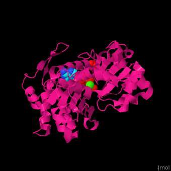
<StructureSection load='3hbt' size='450' side='right' scene='43/430015/Cv/3' caption='Native G-actin with methyl-histidine complex with ATP, sulfate and Ca+2 ion (green) [[3hbt]]'>
{{STRUCTURE_3hbt|  PDB=3hbt  | SIZE=400| SCENE=Actin/Cv/1 |right|CAPTION=Native G-actin with methyl-histidine complex with ATP, sulfate and Ca+2 ion [[3hbt]] }}


== Function ==


[[Actin]] is a protein found in all eukaryotic cells.<ref>PMID:11474115</ref>  It is the monomer of two types of filaments: microfilaments which are part of the cytoskeleton and thin filaments which are part of muscles.  Three isoforms of actin are identified: α (Aa) (or G-actin) found in muscle tissue, β (Ab) and γ (Ag) actins  are components of the cytoskeleton.  F-actin is Aa bound to ATP.  For more details see:<br /> *[[F-actin]]<br /> *[[Non-polymerizable monomeric actin]].<br /> <scene name='Actin/Cv/2'>Click here to see the difference between 2 conformations of bovine Ag actin</scene> (PDB entries [[1hlu]] and [[2btf]]; morph was taken from [http://molmovdb.org/cgi-bin/movie.cgi Gallery of Morphs] of the [http://molmovdb.org Yale Morph Server]).  Actin participates in muscle contraction, cell motility, cell division and cytokinesis.  Actin associated with myosin is responsible for most cell movements.
*'''α actin''' is found exclusively in muscle fibres.<br />
*'''β actin''' is required for early embryonic development<ref>PMID:21900491</ref>.<br />
*'''γ actin''' is required for cytoskeletal maintenance<ref>PMID:19497859</ref>.


See also [[Actin Protein (Hebrew)]]


== Disease ==


Mutations in α-actin found in skeletal striated muscles can cause myopathy.  Mutations in α-actin found in smooth muscles can cause thoracic aortic aneurism. Mutations in α-actin found in heart muscles can cause heart malfunctioning.


== Structural highlights ==


<scene name='43/430015/Cv/10'>Actin binds ATP</scene> in a cleft. Water molecules are shown as red spheres. <scene name='43/430015/Cv/13'>ATP and Ca2+ ion are located in cleft</scene>.  <scene name='43/430015/Cv/11'>Click here to see Ca2+ ion coordination site</scene>.<ref>PMID:20540085</ref> It changes its conformation upon hydrolysis of its bound ATP to ADP.  Actin filaments are polar.  They are formed with all monomers having their clefts pointing in the same direction.


[[Actin]] is a protein found in all eukaryotic cells.  It is the monomer of two types of filaments: microfilaments which are part of the cytoskeleton and thin filaments which are part of muscles.  Three isoforms of actin are identified: α (Aa) (or G-actin) found in muscle tissue, β (Ab) and γ (Ag) actins  are components of the cytoskeleton.  F-actin is Aa bound to ATP. The images at the left and at the right correspond to one representative Actin, ''i.e.'' the crystal structure of rabbit Native G-actin ([[3hbt]]).  For more details see:<br /> *[[F-actin]]<br /> *[[Non-polymerizable monomeric actin]].<br /> <scene name='Actin/Cv/2'>Click here to see the difference between 2 conformations of bovine Ag actin</scene> (PDB entries [[1hlu]] and [[2btf]]; morph was taken from [http://molmovdb.org/cgi-bin/movie.cgi Gallery of Morphs] of the [http://molmovdb.org Yale Morph Server]).
<br />
<br />
<br />
<br />
<br />
== 3D Structures of Actin ==
[[Actin 3D structures]]


Updated on {{REVISIONDAY2}}-{{MONTHNAME|{{REVISIONMONTH}}}}-{{REVISIONYEAR}}
</StructureSection>
 
=== α actin 1 (Aa) ===
 
[[2w49]] – cAa +troponin C+ troponin T+ troponin I+ tropomyosin α-1 – chicken<br />
[[1mdu]] – cAa+hGelsolin<br />
[[3hbt]], [[2zwh]], [[1nwk]], [[1j6z]], [[3g37]], [[2y83]] – rAa – rabbit<br />
[[4a7f]], [[4a7h]], [[4a7l]] – rAa + tropomyosin α-1 + myosin heavy chain (mutant) – Cryo EM<br />
[[2q1n]], [[2q31]], [[2a5x]], [[1rdw]], [[1rfq]], [[1lcu]] – rAa – dimer<br />
[[3mfp]], [[4a7n]] – rAa – Cryo EM<br />
[[2q36]], [[1qz5]] – rAa+kabiramide C – dimer<br />
[[2hmp]] – rAa – protease cleaved<br />
[[2zwh]], [[2gwj]], [[2gwk]] – rAa-ATP<br />
[[2vyp]] – rAa+myxobacterial rhizopodin<br />
[[2v51]], [[2v52]], [[2yje]], [[2yjf]] – rAa+mMKL/Myocardin-like protein – mouse (m)<br />
[[2vcp]] – rAa+ neural wiskott-aldrich syndrome protein (hWASP) – human (h)<br />
[[2a3z]], [[2a40]], [[2a41]] – rAa+cDNase I+domains of hWASP – cow (c)<br />
[[2ff3]] - rAa+ hGelsolin +domains of hWASP <br />
[[2a42]] – rAa+cDNase I<br />
[[4eah]] – rAa + formin-like protein 3 FH2 domain<br />
[[3daw]] – rAa+mTwinfilin-1<br />
[[3buz]] – rAa+Iota toxin component IA<br />
[[3b5u]] – rAa filament – fiber diffraction<br />
[[1eqy]], [[3cjb]], [[1p8z]], [[3ffk]] – rAa+hGelsolin<br />
[[1rgi]] - rAa+hoGelsolin – horse (ho)<br />
[[1t44]] - rAa+hGelsolin:mThymosin β-4 <br />
[[3u8x]] - rAa+DmThymosin β-4 - ''Drosophila melanogaster''<br />
[[1esv]] - rAa+hGelsolin+ latrunculin A<br />
[[3cjc]] - rAa+hGelsolin+cDNase I<br />
[[2d1k]] - rAa+hWH2 domain of MIN+cDNase I<br />
[[2pbd]] – rAa+hProfilin-1<br />
[[2pav]] - rAa+hProfilin-1+hVASP<br />
[[2q97]] – rAa+toxofilin – ''Toxoplasma gondii''<br />
[[2q0r]] – rAa+pectenotoxin-2<br />
[[2q0u]] - rAa+pectenotoxin-2+latrunculin B<br />
[[1ijj]] - rAa+latrunculin A<br />
[[2fxu]] – rAa+bistramide A<br />
[[1wua]] – rAa+alpyronine A<br />
[[2asm]], [[2asp]] – rAa+reidispongiolide<br />
[[2aso]] – rAa+sphinxolide B<br />
[[1yxq]] – rAa+swinholide A<br />
[[1y64]] – rAa+yBNI1 domain FH2 - yeast<br />
[[1sqk]] – rAa+DmCiboulot <br />
[[3sjh]], [[3u9d]], [[3u9z]] - rAa+DmCiboulot thymosin β-4 domain<br />
[[3mn5]] – rAa+ DmSpire WH2 domain<br />
[[2ff6]] - rAa+DmCiboulot+hGelsolin <br />
[[1s22]] – rAa+ulapualide A<br />
[[1qz6]] – rAa+jaspisamide A<br />
[[1lot]], [[1kxp]], [[1ma9]] – rAa+hVitamin D binding protein<br />
[[3m1f]] – rAa+VOPL WH2 domain<br />
[[3m3n]] – rAa+mN-WASP<br />
[[3m6g]] – rAa+lobophorolide<br />
[[3tpq]] – rAa + MAL RPEL domain<br />
[[3ue5]] – rAa + spire domain D<br />
[[4gy2]] – rAa + ι toxin component IA <br />
[[4h0t]] – rAa + ι toxin component IA + ADPR<br />
[[4h03]], [[4h0v]], [[4h0x]], [[4h0y]] – rAa + ι toxin component IA + NAD<br />
[[4jhd]] – DmAa + protein cordon-bleu WH2 domain<br />
 
=== β actin (Ab) ===
 
[[3lue]] – hAb+hAlpha-actinin <br />
[[3byh]] – hAb+hFimbrin ABD2 – Cryo EM<br />
[[2oan]] – cAb<br />
[[3j0s]] – cAb + cofilin 2<br />
[[3u4l]], [[3ub5]] – Ab + profilin-1 - bovine
 
=== γ 1 actin (Ag) ===


[[1atn]] – rAg+cDNase I<br />
[[3chw]] – Major DdAg+hProfilin+poly-Pro repeat – ''Dictyostelium discoideum''<br />
[[3ci5]], [[3cip]], [[1nmd]] - Major DdAg+hGelsolin<br />
[[3a5l]], [[3a5m]], [[3a5n]], [[3a5o]] - Major DdAg+hGelsolin (mutant)<br />
[[1c0g]], [[1dej]] – DdAg:tetrahymenatA+hGelsolin<br />
[[1nm1]] - DdAg+hGelsolin+Mg-ATP<br />
[[1nlv]], [[1c0f]] – DdAg+hGelsolin<br />
[[1d4x]] – Ag 1/3+hGelsolin – ''C. elegans''<br />
[[1h1v]] – hAg+rGelsolin<br />
[[1hlu]], [[2btf]] – cA+cProfilin<br />
[[1o1g]], [[1o1a]], [[1o1b]], [[1o1c]], [[1o1d]], [[1o1e]], [[1o1f]], [[1o18]], [[1o19]], [[1mvw]], [[1m8q]] –cAg+cMyosin II - tomography<br />
[[3eks]] – DmAg 5c (mutant)<br />
[[3eku]] – DmAg 5c (mutant)+cytochalasin D<br />
[[3el2]], [[2hf4]] - DmAg 5c (mutant)+Ca-ATP<br />
[[2hf3]] - DmAg 5c (mutant)+Ca-ADP<br />
[[3w3d]] – cAg + DNase I<br />
[[3b63]] – actin filament – ''Limulus polyphemus''<br />
[[1yvn]] – yA (mutant)+hGelsolin <br />
[[1yag]] - yA+hGelsolin<br />
[[3mmv]] – DmA-5C+spire WH2 domain<br />
[[3mn6]], [[3mn7]], [[3mn9]] - DmA-5C (mutant)+spire<br />
[[4efh]] – A + spire domains C,D – ''Acanthamoeba castellanii''


== Reference ==
<references/>
[[Category:Topic Page]]
[[Category:Topic Page]]

Latest revision as of 11:40, 2 January 2025


Function

Actin is a protein found in all eukaryotic cells.[1] It is the monomer of two types of filaments: microfilaments which are part of the cytoskeleton and thin filaments which are part of muscles. Three isoforms of actin are identified: α (Aa) (or G-actin) found in muscle tissue, β (Ab) and γ (Ag) actins are components of the cytoskeleton. F-actin is Aa bound to ATP. For more details see:
*F-actin
*Non-polymerizable monomeric actin.
(PDB entries 1hlu and 2btf; morph was taken from Gallery of Morphs of the Yale Morph Server). Actin participates in muscle contraction, cell motility, cell division and cytokinesis. Actin associated with myosin is responsible for most cell movements.

  • α actin is found exclusively in muscle fibres.
  • β actin is required for early embryonic development[2].
  • γ actin is required for cytoskeletal maintenance[3].

See also Actin Protein (Hebrew)

Disease

Mutations in α-actin found in skeletal striated muscles can cause myopathy. Mutations in α-actin found in smooth muscles can cause thoracic aortic aneurism. Mutations in α-actin found in heart muscles can cause heart malfunctioning.

Structural highlights

in a cleft. Water molecules are shown as red spheres. . .[4] It changes its conformation upon hydrolysis of its bound ATP to ADP. Actin filaments are polar. They are formed with all monomers having their clefts pointing in the same direction.

3D Structures of Actin

Actin 3D structures


Native G-actin with methyl-histidine complex with ATP, sulfate and Ca+2 ion (green) 3hbt

Drag the structure with the mouse to rotate


ReferenceReference

  1. Otterbein LR, Graceffa P, Dominguez R. The crystal structure of uncomplexed actin in the ADP state. Science. 2001 Jul 27;293(5530):708-11. PMID:11474115 doi:10.1126/science.1059700
  2. Bunnell TM, Burbach BJ, Shimizu Y, Ervasti JM. β-Actin specifically controls cell growth, migration, and the G-actin pool. Mol Biol Cell. 2011 Nov;22(21):4047-58. PMID:21900491 doi:10.1091/mbc.E11-06-0582
  3. Belyantseva IA, Perrin BJ, Sonnemann KJ, Zhu M, Stepanyan R, McGee J, Frolenkov GI, Walsh EJ, Friderici KH, Friedman TB, Ervasti JM. Gamma-actin is required for cytoskeletal maintenance but not development. Proc Natl Acad Sci U S A. 2009 Jun 16;106(24):9703-8. PMID:19497859 doi:10.1073/pnas.0900221106
  4. Wang H, Robinson RC, Burtnick LD. The structure of native G-actin. Cytoskeleton (Hoboken). 2010 Jul;67(7):456-65. PMID:20540085 doi:10.1002/cm.20458

Proteopedia Page Contributors and Editors (what is this?)Proteopedia Page Contributors and Editors (what is this?)

Alexander Berchansky, Michal Harel, Joel L. Sussman