1d0g: Difference between revisions
No edit summary |
No edit summary |
||
| (14 intermediate revisions by the same user not shown) | |||
| Line 1: | Line 1: | ||
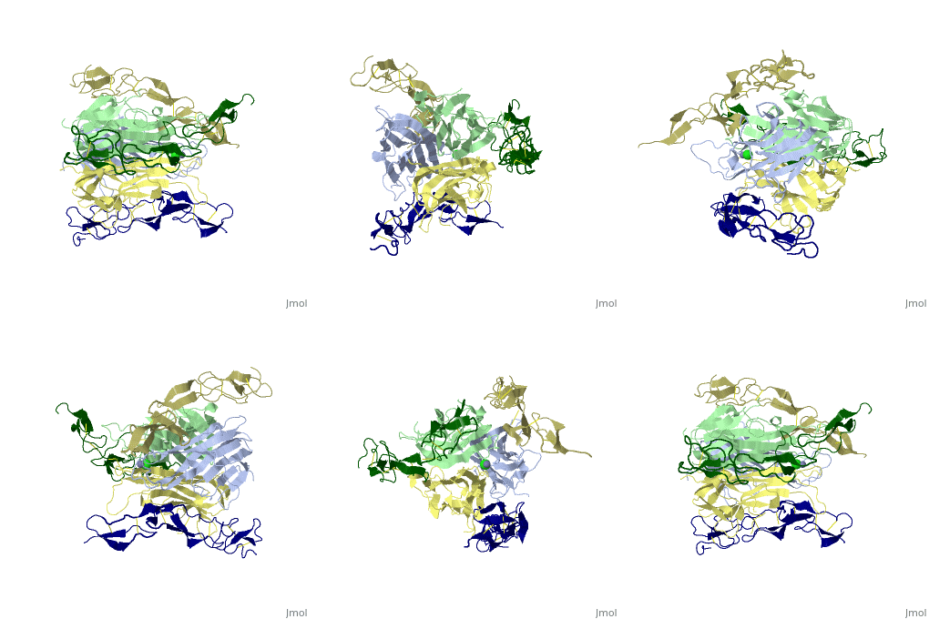
==CRYSTAL STRUCTURE OF DEATH RECEPTOR 5 (DR5) BOUND TO APO2L/TRAIL== | |||
<StructureSection load='1d0g' size='340' side='right'caption='[[1d0g]], [[Resolution|resolution]] 2.40Å' scene=''> | |||
== Structural highlights == | |||
<table><tr><td colspan='2'>[[1d0g]] is a 6 chain structure with sequence from [https://en.wikipedia.org/wiki/Homo_sapiens Homo sapiens]. Full crystallographic information is available from [http://oca.weizmann.ac.il/oca-bin/ocashort?id=1D0G OCA]. For a <b>guided tour on the structure components</b> use [https://proteopedia.org/fgij/fg.htm?mol=1D0G FirstGlance]. <br> | |||
</td></tr><tr id='method'><td class="sblockLbl"><b>[[Empirical_models|Method:]]</b></td><td class="sblockDat" id="methodDat">X-ray diffraction, [[Resolution|Resolution]] 2.4Å</td></tr> | |||
<tr id='ligand'><td class="sblockLbl"><b>[[Ligand|Ligands:]]</b></td><td class="sblockDat" id="ligandDat"><scene name='pdbligand=CL:CHLORIDE+ION'>CL</scene>, <scene name='pdbligand=ZN:ZINC+ION'>ZN</scene></td></tr> | |||
<tr id='resources'><td class="sblockLbl"><b>Resources:</b></td><td class="sblockDat"><span class='plainlinks'>[https://proteopedia.org/fgij/fg.htm?mol=1d0g FirstGlance], [http://oca.weizmann.ac.il/oca-bin/ocaids?id=1d0g OCA], [https://pdbe.org/1d0g PDBe], [https://www.rcsb.org/pdb/explore.do?structureId=1d0g RCSB], [https://www.ebi.ac.uk/pdbsum/1d0g PDBsum], [https://prosat.h-its.org/prosat/prosatexe?pdbcode=1d0g ProSAT]</span></td></tr> | |||
</table> | |||
== Disease == | |||
[https://www.uniprot.org/uniprot/TR10B_HUMAN TR10B_HUMAN] Defects in TNFRSF10B may be a cause of head and neck squamous cell carcinomas (HNSCC) [MIM:[https://omim.org/entry/275355 275355]; also known as squamous cell carcinoma of the head and neck. | |||
== Function == | |||
[https://www.uniprot.org/uniprot/TR10B_HUMAN TR10B_HUMAN] Receptor for the cytotoxic ligand TNFSF10/TRAIL. The adapter molecule FADD recruits caspase-8 to the activated receptor. The resulting death-inducing signaling complex (DISC) performs caspase-8 proteolytic activation which initiates the subsequent cascade of caspases (aspartate-specific cysteine proteases) mediating apoptosis. Promotes the activation of NF-kappa-B. Essential for ER stress-induced apoptosis.<ref>PMID:15322075</ref> | |||
== Evolutionary Conservation == | |||
[[Image:Consurf_key_small.gif|200px|right]] | |||
Check<jmol> | |||
<jmolCheckbox> | |||
<scriptWhenChecked>; select protein; define ~consurf_to_do selected; consurf_initial_scene = true; script "/wiki/ConSurf/d0/1d0g_consurf.spt"</scriptWhenChecked> | |||
<scriptWhenUnchecked>script /wiki/extensions/Proteopedia/spt/initialview03.spt</scriptWhenUnchecked> | |||
<text>to colour the structure by Evolutionary Conservation</text> | |||
</jmolCheckbox> | |||
</jmol>, as determined by [http://consurfdb.tau.ac.il/ ConSurfDB]. You may read the [[Conservation%2C_Evolutionary|explanation]] of the method and the full data available from [http://bental.tau.ac.il/new_ConSurfDB/main_output.php?pdb_ID=1d0g ConSurf]. | |||
<div style="clear:both"></div> | |||
<div style="background-color:#fffaf0;"> | |||
== Publication Abstract from PubMed == | |||
Formation of a complex between Apo2L (also called TRAIL) and its signaling receptors, DR4 and DR5, triggers apoptosis by inducing the oligomerization of intracellular death domains. We report the crystal structure of the complex between Apo2L and the ectodomain of DR5. The structure shows three elongated receptors snuggled into long crevices between pairs of monomers of the homotrimeric ligand. The interface is divided into two distinct patches, one near the bottom of the complex close to the receptor cell surface and one near the top. Both patches contain residues that are critical for high-affinity binding. A comparison to the structure of the lymphotoxin-receptor complex suggests general principles of binding and specificity for ligand recognition in the TNF receptor superfamily. | |||
Triggering cell death: the crystal structure of Apo2L/TRAIL in a complex with death receptor 5.,Hymowitz SG, Christinger HW, Fuh G, Ultsch M, O'Connell M, Kelley RF, Ashkenazi A, de Vos AM Mol Cell. 1999 Oct;4(4):563-71. PMID:10549288<ref>PMID:10549288</ref> | |||
From MEDLINE®/PubMed®, a database of the U.S. National Library of Medicine.<br> | |||
</div> | |||
<div class="pdbe-citations 1d0g" style="background-color:#fffaf0;"></div> | |||
== | ==See Also== | ||
*[[TRAIL|TRAIL]] | |||
*[[Tumor necrosis factor receptor 3D structures|Tumor necrosis factor receptor 3D structures]] | |||
== References == | |||
<references/> | |||
__TOC__ | |||
</StructureSection> | |||
[[Category: Homo sapiens]] | [[Category: Homo sapiens]] | ||
[[Category: | [[Category: Large Structures]] | ||
[[Category: Ashkenazi | [[Category: Ashkenazi A]] | ||
[[Category: Christinger | [[Category: Christinger HW]] | ||
[[Category: Fuh G]] | |||
[[Category: Fuh | [[Category: Hymowitz SG]] | ||
[[Category: Hymowitz | [[Category: Kelley RF]] | ||
[[Category: Kelley | [[Category: O'Connell MP]] | ||
[[Category: | [[Category: De Vos AM]] | ||
[[Category: | |||
Latest revision as of 07:26, 17 October 2024
CRYSTAL STRUCTURE OF DEATH RECEPTOR 5 (DR5) BOUND TO APO2L/TRAILCRYSTAL STRUCTURE OF DEATH RECEPTOR 5 (DR5) BOUND TO APO2L/TRAIL
Structural highlights
DiseaseTR10B_HUMAN Defects in TNFRSF10B may be a cause of head and neck squamous cell carcinomas (HNSCC) [MIM:275355; also known as squamous cell carcinoma of the head and neck. FunctionTR10B_HUMAN Receptor for the cytotoxic ligand TNFSF10/TRAIL. The adapter molecule FADD recruits caspase-8 to the activated receptor. The resulting death-inducing signaling complex (DISC) performs caspase-8 proteolytic activation which initiates the subsequent cascade of caspases (aspartate-specific cysteine proteases) mediating apoptosis. Promotes the activation of NF-kappa-B. Essential for ER stress-induced apoptosis.[1] Evolutionary Conservation Check, as determined by ConSurfDB. You may read the explanation of the method and the full data available from ConSurf. Publication Abstract from PubMedFormation of a complex between Apo2L (also called TRAIL) and its signaling receptors, DR4 and DR5, triggers apoptosis by inducing the oligomerization of intracellular death domains. We report the crystal structure of the complex between Apo2L and the ectodomain of DR5. The structure shows three elongated receptors snuggled into long crevices between pairs of monomers of the homotrimeric ligand. The interface is divided into two distinct patches, one near the bottom of the complex close to the receptor cell surface and one near the top. Both patches contain residues that are critical for high-affinity binding. A comparison to the structure of the lymphotoxin-receptor complex suggests general principles of binding and specificity for ligand recognition in the TNF receptor superfamily. Triggering cell death: the crystal structure of Apo2L/TRAIL in a complex with death receptor 5.,Hymowitz SG, Christinger HW, Fuh G, Ultsch M, O'Connell M, Kelley RF, Ashkenazi A, de Vos AM Mol Cell. 1999 Oct;4(4):563-71. PMID:10549288[2] From MEDLINE®/PubMed®, a database of the U.S. National Library of Medicine. See AlsoReferences
|
| ||||||||||||||||||
