1dd3: Difference between revisions
Jump to navigation
Jump to search
No edit summary |
No edit summary |
||
| (17 intermediate revisions by the same user not shown) | |||
| Line 1: | Line 1: | ||
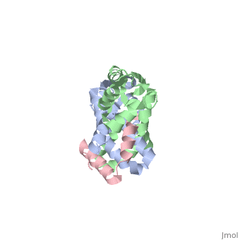
==CRYSTAL STRUCTURE OF RIBOSOMAL PROTEIN L12 FROM THERMOTOGA MARITIMA== | |||
<StructureSection load='1dd3' size='340' side='right'caption='[[1dd3]], [[Resolution|resolution]] 2.00Å' scene=''> | |||
== Structural highlights == | |||
<table><tr><td colspan='2'>[[1dd3]] is a 4 chain structure with sequence from [https://en.wikipedia.org/wiki/Thermotoga_maritima Thermotoga maritima]. Full crystallographic information is available from [http://oca.weizmann.ac.il/oca-bin/ocashort?id=1DD3 OCA]. For a <b>guided tour on the structure components</b> use [https://proteopedia.org/fgij/fg.htm?mol=1DD3 FirstGlance]. <br> | |||
</td></tr><tr id='method'><td class="sblockLbl"><b>[[Empirical_models|Method:]]</b></td><td class="sblockDat" id="methodDat">X-ray diffraction, [[Resolution|Resolution]] 2Å</td></tr> | |||
<tr id='resources'><td class="sblockLbl"><b>Resources:</b></td><td class="sblockDat"><span class='plainlinks'>[https://proteopedia.org/fgij/fg.htm?mol=1dd3 FirstGlance], [http://oca.weizmann.ac.il/oca-bin/ocaids?id=1dd3 OCA], [https://pdbe.org/1dd3 PDBe], [https://www.rcsb.org/pdb/explore.do?structureId=1dd3 RCSB], [https://www.ebi.ac.uk/pdbsum/1dd3 PDBsum], [https://prosat.h-its.org/prosat/prosatexe?pdbcode=1dd3 ProSAT]</span></td></tr> | |||
</table> | |||
== Function == | |||
[https://www.uniprot.org/uniprot/RL7_THEMA RL7_THEMA] Seems to be the binding site for several of the factors involved in protein synthesis and appears to be essential for accurate translation. | |||
== Evolutionary Conservation == | |||
[[Image:Consurf_key_small.gif|200px|right]] | |||
Check<jmol> | |||
<jmolCheckbox> | |||
<scriptWhenChecked>; select protein; define ~consurf_to_do selected; consurf_initial_scene = true; script "/wiki/ConSurf/dd/1dd3_consurf.spt"</scriptWhenChecked> | |||
<scriptWhenUnchecked>script /wiki/extensions/Proteopedia/spt/initialview01.spt</scriptWhenUnchecked> | |||
<text>to colour the structure by Evolutionary Conservation</text> | |||
</jmolCheckbox> | |||
</jmol>, as determined by [http://consurfdb.tau.ac.il/ ConSurfDB]. You may read the [[Conservation%2C_Evolutionary|explanation]] of the method and the full data available from [http://bental.tau.ac.il/new_ConSurfDB/main_output.php?pdb_ID=1dd3 ConSurf]. | |||
<div style="clear:both"></div> | |||
==See Also== | |||
*[[Ribosomal protein L7/L12|Ribosomal protein L7/L12]] | |||
__TOC__ | |||
== | </StructureSection> | ||
[[Category: Large Structures]] | |||
[[Category: | |||
[[Category: Thermotoga maritima]] | [[Category: Thermotoga maritima]] | ||
[[Category: Bartunik | [[Category: Bartunik HD]] | ||
[[Category: Bourenkov | [[Category: Bourenkov GP]] | ||
[[Category: Huber | [[Category: Huber R]] | ||
[[Category: Wahl | [[Category: Wahl MC]] | ||
Latest revision as of 09:52, 7 February 2024
CRYSTAL STRUCTURE OF RIBOSOMAL PROTEIN L12 FROM THERMOTOGA MARITIMACRYSTAL STRUCTURE OF RIBOSOMAL PROTEIN L12 FROM THERMOTOGA MARITIMA
Structural highlights
FunctionRL7_THEMA Seems to be the binding site for several of the factors involved in protein synthesis and appears to be essential for accurate translation. Evolutionary Conservation Check, as determined by ConSurfDB. You may read the explanation of the method and the full data available from ConSurf. See Also |
| ||||||||||||||||