MDM2
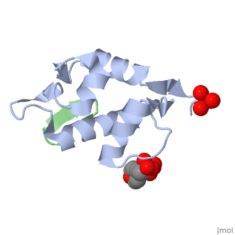
FunctionThe MDM2 or E3 ubiquitin-protein ligase MDM2 oncoprotein is a cellular inhibitor of the p53 tumor suppressor in that it can bind the transactivation domain of p53 and downregulate its ability to activate transcription. In certain cancers, MDM2 amplification is a common event and contributes to the inactivation of p53. The crystal structure of the 109-residue amino-terminal domain of MDM2 bound to 109-residue transactivation domain peptide of p53 revealed that MDM2 has a on which the p53 peptide binds as an amphipathic alpha helix. The interface relies on the steric complementarity between the MDM2 cleft and the hydrophobic face of the p53 alpha helix and, in particular, on a triad of p53 amino acids-Phe19, Trp23, and Leu26-which insert the MDM2 cleft. These same p53 residues are also involved in transactivation, supporting the hypothesis that MDM2 inactivates p53 by concealing its transactivation domain. The structure also suggests that the amphipathic alpha helix may be a common structural motif in the binding of a diverse family of transactivation factors to the TATA-binding protein-associated factors.[1] Role in CancerMDM2 is an active inhibitor of the p53 anti-tumorgenesis protein. When MDM2 is atached p53 is unable to release transcription factors that code for apoptosis or halting of the cell cycle. The full cell signaling pathway is very complex, but MDM2 overproduction has been shown to be connected to cancer activity.[2] Genetic amplification of MDM2 has been shown in breast, glioma, pancreaticadenocarcinoma, leucemias, Hodgkin's and nonhodgkin's lymphomas and other various cancers.[2] This broad observation outlines the importance of determining MDM2's role in tumer-genesis and how the process can be treated. See also Proteins involved in cancer. Experimental ResearchEarly work in the field of MDM2 overproduction in tumor cells was done by Raphael E. Pollok et. al. This research consisted of tests done with cancerous tumor tissue and analogous tissue. DNA amplification, mRNA creation, and protein creation were all areas of interest in determining what mechanism was being utilized for the overexertion of MDM2. The amplification of the DNA would show a DNA based increase in coding for the protein. An excess of mRNA would show the increase in protein was due to increased transcription factors for MDM2. The last possibility would be that the overproduction of MDM2 was an error in translation and only protein production was to blame. These factors were tested using Southern, Northern, and Western plots to analyze DNA, mRNA, and protein production respectively. These tests use targeting antigens to test for the presence of desired proteins. The results of these tests reveled the genetic amplification of the DNA was to blame for the increased of MDM2. Also evidence that p53 protein mutations affect MDM2 were found. These results are of great use to focus treatment possibilities and further research.[2] 3D structures of MDM2
|
| ||||||||||
Additional ResourcesAdditional Resources
For additional information, see: Cancer
ReferenceReference
- ↑ Kussie PH, Gorina S, Marechal V, Elenbaas B, Moreau J, Levine AJ, Pavletich NP. Structure of the MDM2 oncoprotein bound to the p53 tumor suppressor transactivation domain. Science. 1996 Nov 8;274(5289):948-53. PMID:8875929
- ↑ 2.0 2.1 2.2 Pollock RE, Lang A, El-Naggar AK, Radinsky R, Hung MC. Enhanced MDM2 Oncoprotein Expression in Soft Tissue Sarcoma: Several Possible Regulatory Mechanisms. Sarcoma. 1997;1(1):23-9. PMID:18521197 doi:10.1080/13577149778443