Horse Liver Alcohol Dehydrogenase
General InformationHorse liver alcohol dehydrogenase (LADH) is found in the liver of the species Equus Caballus (horse). It is a dimeric zinc-dependant protein, with a broad substrate specificity, being active on a variety of primary, secondary, branched, and cyclic alcohols.[1] The enzymatic mechanism and structures of various complexes have been extensively studied.[2][3][4] The catalytic reaction is ordered, with NAD+ or NADH binding to free enzyme when primary alcohols and aldehydes, such as ethanol and acetaldehyde or benzyl alcohol and benzaldehyde, are the substrates.[1]The enzyme has a molecular weight of 80 000 and is a dimmer of two identical subunits, each of which has a coenzyme binding and a catalytic domain. The coenzyme-binding domains have structures similar to those found in several other NAD-dependent dehydrogenases.[1] Protein StructureHorse liver alcohol dehydrogenase is a homodimeric protein. Each monomer contains 374 amino acids and has a mass of 40 kDa, with each monomer also containing a catalytic and coenzyme binding domain.[1] The coenzyme-binding domains have structures similar to those found in several other NAD-dependent dehydrogenases,[1]and they form a 12-stranded beta-pleated sheet structure that makes up the central core of the dimer.[2] Through crystal structure analysis of horse liver alcohol dehydrogenase, which has been extended to 2.4 Å resolution, electron density maps of the apoenzyme have determined the positions of the 374 amino acids in the polypeptide chain of each subunit.[5] The coenzyme binding domain of the subunit comprises of residues 176 to 318. Fourty-five percent of these residues are helical and 32% are in the central six-stranded pleated sheet structure.[5]The positions and orientations of the helices with respect to the pleated sheet indicate a possible folding mechanism for this part of the subunit structure.[5] The catalytic domain is mainly built up from three distinct antiparallel pleated-sheet regions. Residues within this domain provide ligands to the catalytic atom; Cys46, His67 and Cys174.[6] An approximate tetrahedral coordination of this zinc is completed by a water molecule or hydroxyl ion depending on the pH. Residues 95 to 113 form a lobe that binds the second zinc atom of the subunit. This zinc is liganded in a distorted tetrahedral arrangement by four sulphur atoms from the cysteine residues 97, 100, 103 and 111. The lobe forms one side of a significant cleft in the enzyme surface suggesting that this region might constitute a second catalytic centre of unknown function.[6]
The two domains of the subunit are separated by a crevice that contains a wide and deep hydrophobic pocket. The catalytic zinc atom is at the bottom of this pocket, with the zinc-bound water molecule projecting out into the pocket. This water molecule is hydrogen-bonded to the side chain of Ser48 which in turn is hydrogen-bonded to His51.[4] The pocket which in all probability is the binding site for the substrate and the nicotinamide moiety of the coenzyme, is lined almost exclusively with hydrophobic side chains.[3][4] The only accessible polar groups in the vicinity of the catalytic centre are Ser48 and Thr178 apart from zinc and the zinc-bound water molecule.[6] From structural determinants by x-ray diffraction it is known that LADH undergoes conformational changes going from an open structure, in which large binding areas for coenzymes and substrates are accessible, towards a closed conformation.[3] However the trigger mechanism for the structural transition is not yet fully understood. A complex pattern has emerged, showing that the structural changes in the LADH depend on coenzyme analogue structure and on which combination of coenzyme and a second ligand is present.[4] Protein FunctionLADH catalyzes the transfer of hydride ion from an alcohol substrate to the NAD+ cofactor, yielding an aldehyde and the reduced cofactor, NADH. The two active sites are in clefts between the coenzyme-binding core and the catalytic domains, and contain a tetracoordinate zinc cations[3],with three of the zinc ions being contributed by the enzyme. In the “open” form of the apoenzyme, the active site is available to solvent and the fourth zinc ion is occupied by water[3]. Substrates bind in a hydrophobic pocket in the cleft with their oxygen atoms ligated to the zinc atoms of the catalytic domains. When coenzymes bind, the active site clefts close up by a rigid body rotation of the catalytic domains toward the coenzyme-binding domains.[1] Once the enzyme adopts the closed form, the substrate and cofactor are sufficiently close to allow direct transfer of a hydride ion from the alcohol secondary carbon to the C4 position of the nicotinamide ring.[3] The specific step which is required for the hydride transfer is the isomerization step.[4] This step requires the rotation of the catalytic domain relative to the coenzyme binding domain, resulting in a narrowing of the interdomain cleft and induces an increase in the hydrophobic environment at the active site.[2]This rearrangement facilitates displacement of water by alcohol with the fourth coordination ligand of the catalytic zinc and places the zinc-bound substrate close to the C4 position of the cofactor’s nicotinamide ring.[2] MutationsCommon mutations within LADH can be used to exploit its hydrogen tunneling ability, and it’s contribution to the hydride transfer step. Such mutations include what is called a F93W mutation, which introduces a bulkier tryptophan replacing Phe93. This increases the product off rate of aldehyde by shrinking the size of the substrate binding pocket through the addition of steric bulk.[3][2] Another mutant design called the V203A, decreases the tunneling activity in the enzyme by altering the active site geometry. The substitution of a valine to a less bulky side chain limits the hydrophobic interaction between the nicotinamide ring and the active site of the enzyme.[3][2] Additional FunctionsWhile LADH is generally associated with the interconversion of alcohols and aldehydes, it has also received some attention for its catalysis of aldehyde. Calculations suggest that it is possible that LADH could catalyze hydration of an aldehyde when hydroxide is ligated to the active site zinc. Molecular dynamics results suggest that the geometry is favorable for nucleophilic attack of zinc bound hydroxide ion on the aldehyde carbonyl carbon.[7] |
| ||||||||||
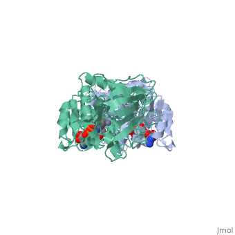
3D structures of horse liver alcohol dehydrogenase3D structures of horse liver alcohol dehydrogenase
ReferencesReferences
- ↑ 1.0 1.1 1.2 1.3 1.4 1.5 Ramaswamy S, Eklund H, Plapp BV. Structures of horse liver alcohol dehydrogenase complexed with NAD+ and substituted benzyl alcohols. Biochemistry. 1994 May 3;33(17):5230-7. PMID:8172897
- ↑ 2.0 2.1 2.2 2.3 2.4 2.5 Bahnson BJ, Colby TD, Chin JK, Goldstein BM, Klinman JP. A link between protein structure and enzyme catalyzed hydrogen tunneling. Proc Natl Acad Sci U S A. 1997 Nov 25;94(24):12797-802. PMID:9371755
- ↑ 3.0 3.1 3.2 3.3 3.4 3.5 3.6 3.7 Colby TD, Bahnson BJ, Chin JK, Klinman JP, Goldstein BM. Active site modifications in a double mutant of liver alcohol dehydrogenase: structural studies of two enzyme-ligand complexes. Biochemistry. 1998 Jun 30;37(26):9295-304. PMID:9649310 doi:10.1021/bi973184b
- ↑ 4.0 4.1 4.2 4.3 4.4 Bahnson BJ, Park DH, Kim K, Plapp BV, Klinman JP. Unmasking of hydrogen tunneling in the horse liver alcohol dehydrogenase reaction by site-directed mutagenesis. Biochemistry. 1993 Jun 1;32(21):5503-7. PMID:8504071
- ↑ 5.0 5.1 5.2 Eklund H, Nordstrom B, Zeppezauer E, Soderlund G, Ohlsson I, Boiwe T, Soderberg BO, Tapia O, Branden CI, Akeson A. Three-dimensional structure of horse liver alcohol dehydrogenase at 2-4 A resolution. J Mol Biol. 1976 Mar 25;102(1):27-59. PMID:178875
- ↑ 6.0 6.1 6.2 Brandt EG, Hellgren M, Brinck T, Bergman T, Edholm O. Molecular dynamics study of zinc binding to cysteines in a peptide mimic of the alcohol dehydrogenase structural zinc site. Phys Chem Chem Phys. 2009 Feb 14;11(6):975-83. Epub 2008 Dec 12. PMID:19177216 doi:10.1039/b815482a
- ↑ Olson LP, Luo J, Almarsson O, Bruice TC. Mechanism of aldehyde oxidation catalyzed by horse liver alcohol dehydrogenase. Biochemistry. 1996 Jul 30;35(30):9782-91. PMID:8703951 doi:10.1021/bi952020x