4cha
STRUCTURE OF ALPHA-*CHYMOTRYPSIN REFINED AT 1.68 ANGSTROMS RESOLUTIONSTRUCTURE OF ALPHA-*CHYMOTRYPSIN REFINED AT 1.68 ANGSTROMS RESOLUTION
Structural highlights
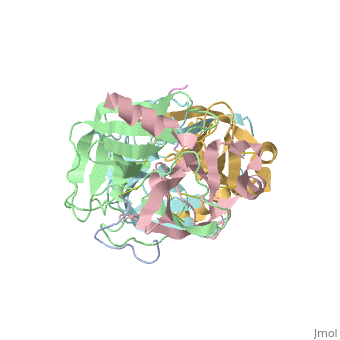
FunctionEvolutionary Conservation Check, as determined by ConSurfDB. You may read the explanation of the method and the full data available from ConSurf. Publication Abstract from PubMedDiffraction data for alpha-chymotrypsin crystals at -10 degrees C were measured at 1.68 A resolution and refined by restrained structure-factor least-squares refinement. The two independent chymotrypsin molecules in the crystallographic asymmetric unit were refined independently. The overall structure of alpha-chymotrypsin is little changed from published co-ordinates. The root-mean-square shift of C alpha co-ordinates is 0.42 A, co-ordinates for the two molecules showing a root-mean-square difference of 0.19 A. Certain regions with high disorder (residues 9 to 14, 73 to 79) remain difficult to interpret and several side-chains are disordered. Some water molecule positions have been changed. The absence of the tosyl group has made a significant difference to the refined structure at the active site. This now agrees closely with other enzymes of the trypsin family that have been refined at high resolution. There is a strong hydrogen bond between N epsilon 2 (His57) and O gamma (Ser195) in the free enzyme, in line with the published description of the charge relay system. Structure of alpha-chymotrypsin refined at 1.68 A resolution.,Tsukada H, Blow DM J Mol Biol. 1985 Aug 20;184(4):703-11. PMID:4046030[1] From MEDLINE®/PubMed®, a database of the U.S. National Library of Medicine. See AlsoReferences |
| ||||||||||||||||