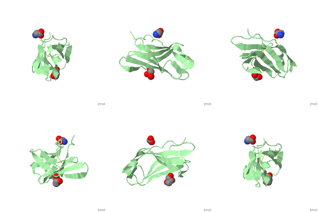
3kgr
Crystal structure of the human leukocyte-associated Ig-like receptor-1 (LAIR-1)Crystal structure of the human leukocyte-associated Ig-like receptor-1 (LAIR-1)
Structural highlights
FunctionLAIR1_HUMAN Functions as an inhibitory receptor that plays a constitutive negative regulatory role on cytolytic function of natural killer (NK) cells, B-cells and T-cells. Activation by Tyr phosphorylation results in recruitment and activation of the phosphatases PTPN6 and PTPN11. It also reduces the increase of intracellular calcium evoked by B-cell receptor ligation. May also play its inhibitory role independently of SH2-containing phosphatases. Modulates cytokine production in CD4+ T-cells, down-regulating IL2 and IFNG production while inducing secretion of transforming growth factor beta. Down-regulates also IgG and IgE production in B-cells as well as IL8, IL10 and TNF secretion. Inhibits proliferation and induces apoptosis in myeloid leukemia cell lines as well as prevents nuclear translocation of NF-kappa-B p65 subunit/RELA and phosphorylation of I-kappa-B alpha/CHUK in these cells. Inhibits the differentiation of peripheral blood precursors towards dendritic cells.[1] [2] [3] [4] [5] [6] [7] [8] [9] [10] Evolutionary Conservation Check, as determined by ConSurfDB. You may read the explanation of the method and the full data available from ConSurf. Publication Abstract from PubMedLeukocyte-associated immunoglobulin-like receptor-1 (LAIR-1), one of the most widely spread immune receptors, attenuates immune cell activation when bound to specific sites in collagen. The collagen-binding domain of LAIR-1 is homologous to that of glycoprotein VI (GPVI), a collagen receptor crucial for platelet activation. Because LAIR-1 and GPVI also display overlapping collagen-binding specificities, a common structural basis for collagen recognition would appear likely. Therefore, it is crucial to gain insight into the molecular interaction of both receptors with their ligand to prevent unwanted cross-reactions during therapeutic intervention. We determined the crystal structure of LAIR-1 and mapped its collagen-binding site by nuclear magnetic resonance (NMR) titrations and mutagenesis. Our data identify R59, E61, and W109 as key residues for collagen interaction. These residues are strictly conserved in LAIR-1 and GPVI alike; however, they are located outside the previously proposed GPVI collagen-binding site. Our data provide evidence for an unanticipated mechanism of collagen recognition common to LAIR-1 and GPVI. This fundamental insight will contribute to the exploration of specific means of intervention in collagen-induced signaling in immunity and hemostasis. Crystal structure and collagen-binding site of immune inhibitory receptor LAIR-1: unexpected implications for collagen binding by platelet receptor GPVI.,Brondijk TH, de Ruiter T, Ballering J, Wienk H, Lebbink RJ, van Ingen H, Boelens R, Farndale RW, Meyaard L, Huizinga EG Blood. 2010 Feb 18;115(7):1364-73. Epub 2009 Dec 10. PMID:20007810[11] From MEDLINE®/PubMed®, a database of the U.S. National Library of Medicine. See AlsoReferences
|
| ||||||||||||||||||
