3fpn
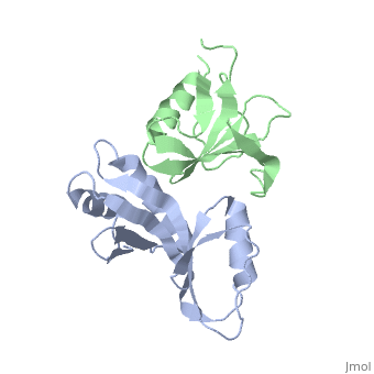
Crystal structure of UvrA-UvrB interaction domainsCrystal structure of UvrA-UvrB interaction domains
Structural highlights
FunctionEvolutionary Conservation Check, as determined by ConSurfDB. You may read the explanation of the method and the full data available from ConSurf. Publication Abstract from PubMedNucleotide excision repair is distinguished from other DNA repair pathways by its ability to process a wide range of structurally unrelated DNA lesions. In bacteria, damage recognition is achieved by the UvrA.UvrB ensemble. Here, we report the structure of the complex between the interaction domains of UvrA and UvrB. These domains are necessary and sufficient for full-length UvrA and UvrB to associate and thereby form the DNA damage-sensing complex of bacterial nucleotide excision repair. The crystal structure and accompanying biochemical analyses suggest a model for the complete damage-sensing complex. A structural model for the damage-sensing complex in bacterial nucleotide excision repair.,Pakotiprapha D, Liu Y, Verdine GL, Jeruzalmi D J Biol Chem. 2009 May 8;284(19):12837-44. Epub 2009 Mar 13. PMID:19287003[1] From MEDLINE®/PubMed®, a database of the U.S. National Library of Medicine. See AlsoReferences
|
| ||||||||||||||||