Sandbox 1
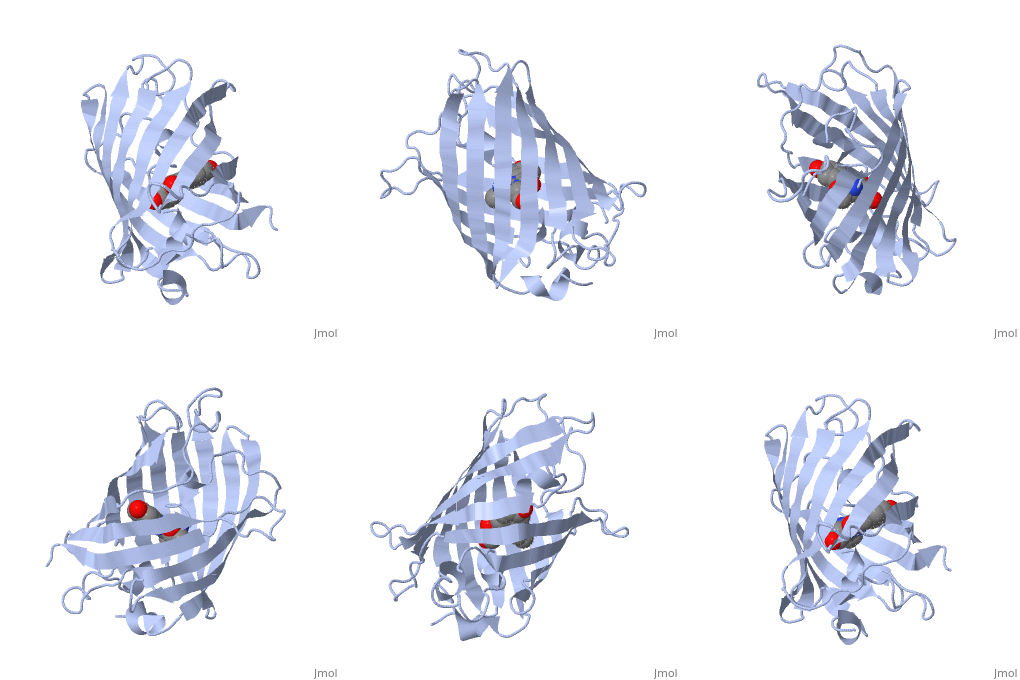
You can take a close look at the chromophore of GFP in the PDB entry 1ema
|
. The backbone of the entire protein is shown here on the left. The protein chain forms a cylindrical can (shown in blue), with one portion of the strand threading straight through the middle (shown in green). The chromophore is found of the can , totally shielded from the surrounding environment.This shielding is essential for the fluorescence. The jostling water molecules would normally rob the chromophore of its energy once it absorbs a photon. But inside the protein, it is protected, releasing the energy instead as a slightly less energetic photon of light. The chromophore (shown in the close-up on the right) forms spontaneously from three amino acids in the protein chain: a glycine, a tyrosine and a threonine (or serine). Notice how the glycine and the threonine have formed a new bond, creating an unusual five-membered ring.
