Titin
Titin (TTN) is the largest known protein. The human TTN contains 34,350 residues. It is responsible for the passive elasticity of muscle. It has 244 domains connected by unstructured regions. The domains unfold when TTN is stretched. (1ya5). Additional details in IntroductionTitin, also known as connectin, is an elastic and approximately 3,6 MDalton large protein which assembles itself to protein filaments. It is made of more than 30000 amino acids and includes 320 protein domains and therefore is known as the largest human protein. It is a part of the sarcomere, the smallest functional unit in the striated muscles. Tintins task in the sarcomere is to center the myosinheads between the actin filaments and to re-establish the unstreched mode. The titin filament together with the actin filament is connected mechanically over the alpha-actinin with the z-disk. This bond is relatively weak and well regulateable, this is a necessary precondition for the unhindered contraction mechanism. Additionally titin binds to telethonin (t-cap) in the region of the z-disk. In the region of the A-band titin is accumulated with its stiff end near the myosin filament and binds to the calcium dependable protease calpain. In the region of the I-band the long polypeptide becomes elastic and can be turned. The assembly of titin differs in the skeletal- and the cardiac muscle, whereby the marginal expandability of the cardiac sarcomer can be explained. The titin filament is not facilitated with the muscle contraction; it just participates in the flexibility and stability of the muscle and helps to define the contraction speed. In addition, titin is responsible for the relaxing mode of the muscle.
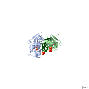
   Titin, the third most abundant and largest known protein with a lenght > 1 μm, is made of more than 300 tandem repeats of immunoglobulin-like (Ig) domains, fibronectin type (FN-III) repeats and from flexible random coil-like PEVK which are rich in proline, glutamate, valine and lysine regions. The insertion of single Lg-domains lead to isoforms of titin that can be found in the cardiac and skeletal muscles. The N-terminal part of titin interacts with some sarcomeric proteins at the Z-disc. These interactions maintain structural assistance for the development and function of myofibrils and also have regulatory influence, such as responding to stretching forces developed in titin. Unique among them, is the structural protein telethonin, which is believed to anchor the ends of two separate titin molecules to the Z-disc. The C-terminal region is a part of the sarcomeric M-line. In picture (Z1Z2 stretched unstretched) one can see the highly schematic view of the tertiary structure elasticity of titin due to bending adjacent protein domains open and closed. In real titin, not every linker may contribute high flexibility, some linkers might be short and stiff. Extensive studies have shown that titin acts in many ways. Titin is believed to be a molecular scaffold in the early growth of myofibrill and that its end is not just an anchor, furthermore they interact with signaling proteins at the Z-line and M-line. It also functions as a molecular spring that dampens sarcomeric extension forces. The I-band region, that forms an elastic connection between the ends of thick filaments and the sarcomeric Z-disc, contains flexible PEVK domains interdigitated between Ig-domain repeats. These random coil PEVK domains are believed to reversibly unfold to permit the extension, like an entropic spring, and to contribute to titin elasticity when weak forces (50 pN) are applied. Titin has soft elasticity regions which are followed by stiffer, nonlinear regions of elasticity. The soft elasticity regions are a generic behavior of random coils formed through PEVK and similar domains and also because of straightening out of titin's multidomain segments. The stiffer elasticity is due to individual domains. Strong forces (160 pN) on titin unravels its secondary structure. Adjacent segments may exhibit a flexibility that leads a titin chain to assume in the relaxed state, a locally compacted shape and a straight shape when under tension. As seen in the image (Z1Z2 / Telethonin complex), the major force enduring component of this complex is an elaborate intermolecular hydrogen bonding network formed across among telethonin and Z1Z2 domains, and not intramolecularly among termini β-strands of individual Z1 or Z2 domains. This shift to a stronger force enduring interface reduces the possibility of unraveling the individual Ig-domains, thus stabilizing the complex. This demonstrates how cross-linking via hydrogen bonds serves as an important mechanism. It functions as a molecular adhesive, increasing the ability of protein complexes to resist against mechanical stress. 3D Structures of Titin3D Structures of TitinUpdated on 03-June-2025 2a38, 6fwx, 6sdb - hTTN residues 1-194 - human 2f8v, 1ya5 - hTTN residues 1-196+telethonin Additional ResourcesSee: Titin Structure & Function for additional information See Alsohttp://www.biophysik.physik.uni-muenchen.de/movies/movies-and-animations |
| ||||||||||
ReferencesReferences
- http://www.ncbi.nlm.nih.gov:80/pmc/articles/PMC1948054/?tool=pmcentrez
- http://www.ks.uiuc.edu/Research/z1z2/
- http://www.ks.uiuc.edu/Research/telethonin/
- http://de.wikipedia.org/wiki/Titin
Created with the participation of Anton Schmidt, Wolfgang Hermann.