1iat
CRYSTAL STRUCTURE OF HUMAN PHOSPHOGLUCOSE ISOMERASE/NEUROLEUKIN/AUTOCRINE MOTILITY FACTOR/MATURATION FACTORCRYSTAL STRUCTURE OF HUMAN PHOSPHOGLUCOSE ISOMERASE/NEUROLEUKIN/AUTOCRINE MOTILITY FACTOR/MATURATION FACTOR
Structural highlights
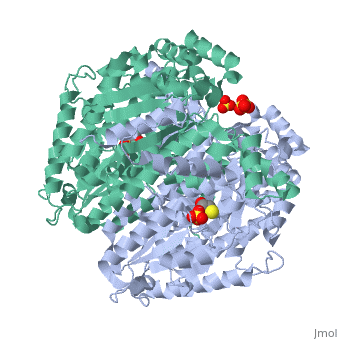
Disease[G6PI_HUMAN] Defects in GPI are the cause of hemolytic anemia non-spherocytic due to glucose phosphate isomerase deficiency (HA-GPID) [MIM:613470]. It is a form of anemia in which there is no abnormal hemoglobin or spherocytosis. It is caused by glucose phosphate isomerase deficiency. Severe GPI deficiency can be associated with hydrops fetalis, immediate neonatal death and neurological impairment. Function[G6PI_HUMAN] Besides it's role as a glycolytic enzyme, mammalian GPI can function as a tumor-secreted cytokine and an angiogenic factor (AMF) that stimulates endothelial cell motility. GPI is also a neurotrophic factor (Neuroleukin) for spinal and sensory neurons.[1] [2] [3] Evolutionary Conservation Check, as determined by ConSurfDB. You may read the explanation of the method and the full data available from ConSurf. Publication Abstract from PubMedPhosphoglucose isomerase (PGI) is a multifunctional protein, which, inside the cell, functions as a housekeeping enzyme of glycolysis and gluconeogenesis and, outside the cell, exerts wholly unrelated cytokine properties. We have determined the structure of human PGI to a resolution of 1.6 A using X-ray crystallography. The structure is highly similar to other PGIs, especially the architecture of the active site. Fortuitous binding of a sulphate molecule from the crystallisation solution has facilitated an accurate description of the substrate phosphate-binding site. Comparison with both native and inhibitor-bound rabbit PGI structures shows that two loops move closer to the active site upon binding inhibitor. Interestingly, the human structure most closely resembles the inhibitor-bound structure, suggesting that binding of the phosphate moiety of the substrate may trigger this conformational change. We suggest a new mechanism for catalysis that uses Glu357 as the base catalyst for the isomerase reaction rather than His388 as proposed previously. The human PGI structure has also provided a detailed framework with which to map mutations associated with non-spherocytic haemolytic anaemia. The crystal structure of human phosphoglucose isomerase at 1.6 A resolution: implications for catalytic mechanism, cytokine activity and haemolytic anaemia.,Read J, Pearce J, Li X, Muirhead H, Chirgwin J, Davies C J Mol Biol. 2001 Jun 1;309(2):447-63. PMID:11371164[4] From MEDLINE®/PubMed®, a database of the U.S. National Library of Medicine. See AlsoReferences
|
| ||||||||||||||||||||