Nitrophorin
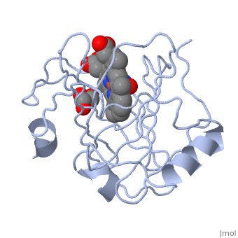
FunctionNitrophorins (NP) are NO transport heme-containing proteins from the saliva of blood-feeding insects which act as vasodilators and anti-platelet agents[1]. The NO is bound to the ferric form of NP with Kds in the micromolar or nanomolar range. Thus upon injection of the saliva into the tissue of the victim, the NO ca dissociate to cause vasodilation and inhibition of platelet aggregation[2]. Structural highlightsThe NPs structures contain a large component of β-sheet structure which is unusual for heme-containing proteins[3]. The NO molecule is bound to the ferric heme opposite the His and Glu residues interacting with it[4]. |
| ||||||||||
3D Structures of deaminase3D Structures of deaminase
Updated on 19-December-2017
ReferencesReferences
- ↑ Andersen JF, Weichsel A, Balfour CA, Champagne DE, Montfort WR. The crystal structure of nitrophorin 4 at 1.5 A resolution: transport of nitric oxide by a lipocalin-based heme protein. Structure. 1998 Oct 15;6(10):1315-27. PMID:9782054
- ↑ Walker FA. Nitric oxide interaction with insect nitrophorins and thoughts on the electron configuration of the {FeNO}6 complex. J Inorg Biochem. 2005 Jan;99(1):216-36. PMID:15598503 doi:http://dx.doi.org/10.1016/j.jinorgbio.2004.10.009
- ↑ Walker FA. Nitric oxide interaction with insect nitrophorins and thoughts on the electron configuration of the {FeNO}6 complex. J Inorg Biochem. 2005 Jan;99(1):216-36. PMID:15598503 doi:http://dx.doi.org/10.1016/j.jinorgbio.2004.10.009
- ↑ Knipp M, Ogata H, Soavi G, Cerullo G, Allegri A, Abbruzzetti S, Bruno S, Viappiani C, Bidon-Chanal A, Luque FJ. Structure and dynamics of the membrane attaching nitric oxide transporter nitrophorin 7. F1000Res. 2015 Feb 13;4:45. doi: 10.12688/f1000research.6060.1. eCollection 2015. PMID:26167269 doi:http://dx.doi.org/10.12688/f1000research.6060.1