IGF1
Insulin-like growth factor 1Insulin-like growth factor 1
Insulin is a very important endocrine protein. Indeed, it is the only hypoglycemic hormone of the human body. This protein is secreted in the beta cells of the Langerhans’ islet in the pancreas and takes part in the glycogenesis. This molecule helps the transportation of glucose into the cells, thus reducing the blood sugar rate, contrary to glucagon. Insulin also has a huge effect on growth and thereby has a lot of structural similarities with its main associated growth factor: IGF-1, aka somatomedin. Both share lots of properties thanks to their 60% similarity. IGF-1 is a peptidic hormone of 69 amino acids which is secreted by the liver. This protein deals with a large scale of regulations, from growth to nutrition and it is even implied in stress response, breeding and longevity.[1]. It is the main actor in primary growth cell control.
HistoryThe role of the pancreas and therefore insulin in diabete was first discovered by Oskar von Minkowski in Strasbourg in 1889. IGF1 was first identified in 1957 by the team of Salmon and Daughaday. In 1976, Rinderknecht and Humbel isolated IGF1 from human cells. The protein was further analysed in 1987 by the team of Dr Mike Davis (Imperial College of London, United Kingdom) and Pr Robin Jokin (INSERM of Toulouse).
Biological structures and interactionsIn the pituitary gland inside the brain, Growth Hormone (GH) is secreted and its release enable transcription of IGF-1 in liver and depending on the nutritional state a paracrine or autocrine activation IGF-1 occurs. Once in the cell, IGF-1 acts as a ligand and can interact with Insulin Receptor protein and Insulin-like Growth Factor Binding Protein. StructuresIGF-1 is a 8.28 kDa protein consisting of 69 amino acids. The coding RNA of this protein creates 4 isoforms differing from each other due to RNA splicing.
Nevertheless, all 4 of them have cysteine residues that are able to make disulfide bond. This enable them to link to IGF-1 receptor.
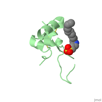
Its secondary structure as shown here is composed of alpha helix and beta strand.
Its 3D structure is :
Stimulating interaction : IGF1 - IGF1RIGF1R is a transmembrane protein receptor. It is composed of two alpha subunits and two tyrosine beta subunits. Both alpha subunits are cysteine-rich region and therefore linked with a disulfide bond. Ligand-binding on alpha subunit induces activation of beta subunit by autophosphorylation. It further leads to activation of the Akt and mTor pathways inside the cell. Due to their homology sequences, the three members of the insulin protein family: IGF1, IGF2 and Insulin can interact with one another of the different receptors. Hence, Insulin binds to IGF1R following the same mechanism and activated the intracellular pathways in the same way IGF1 does. Therefore, the various concentration of the insulin proteins regulates the cell activity in different context, for instance in excess of glucose or lack of Growth Hormone.
Inhibiting interaction : IGF1 - IGFBPIGF binding proteins (IGFBPs) weight 24 to 45 kDa. All six IGFBPs share 50% homology with each other and have binding affinities at the same order of magnitude for IGF-I and IGF-II but have greater affinities then IGF-1 for its receptor. Once IGF1 is bound to Insulin-like Growth Binding Protein (IGFBP), IGF1 cannot be linked to IGF1R any longer. Therefore, increases in serum levels of this IGFBP result in a decrease of IGF-1 activity thus inhibiting the cellular pathways. The IGFBPs help to lengthen the half-life of circulating IGFs in all tissues. That is why approximately 98% of IGF-1 exists as complexed form with one of the six different IGFBP. IGFBP-3, the most abundant protein, accounts for 80% of all IGF binding. Inside the liver, this mechanism is responsible for positive feedback, more precisely it allows growth hormone to continuously act upon the liver to produce more IGF-1.
MetabolismBiological contextRelated diseasesMutations,... Applications |
| ||||||||||||||||

ReferencesReferences
- ↑ Patrick Jouandin. Rôle de la voie de signalisation Insuline dans le couplage des informations nutritionnelles et développementales au cours de l'ovogenèse chez la drosophile. Sciences agricoles.Université Nice Sophia Antipolis, 2013. Français.<NNT : 2013NICE4102>.<tel-00932409>