User:Nikhil Malvankar/Geobacter pilus
Interactive 3D Complement in Proteopedia
 |
![]()
Structural basis for metallic-like conductivity in microbial nanowires.
Nikhil S. Malvankar, Madeline Vargas, Kelly Nevin, Pier-Luc Tremblay, Kenneth Evans-Lutterodt, Dmytro Nykypanchuk, Eric Martz, Mark T. Tuominen, Derek R. Lovley. mBio 6(2):e00084-15 (2015). (doi:10.1128/mBio.00084-15)
Molecular TourMolecular Tour
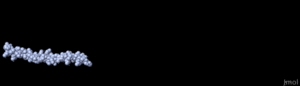
Geobacter Pilin MonomersTwo pilin monomer models () were utilized. First, we made a homology model templated on the X-ray crystallographic structure of the pilin of Pseudomonas aeruginosa[1]. This model is colored in amino-to-carboxy rainbow coloring:
Second, we utilized model 5 of the NMR ensemble of Geobacter sulfurreducens pilin[2]. NMR model 5 is colored magenta. Amino acid sidechains are shown only for aromatics. The models are virtually identical for residues 1-32[3], except for the differing sidechain rotamer positions of Phe1. The models are similarly alpha helical from residues 33-51. Residues 52-61 vary in conformation in the NMR ensemble, while for residues 52-60 in the homology model, the template is unlikely to be appropriate since Pseudomonas has a substantial domain from 52-144 while Geobacter pilin ends at 61. Thus, while one can be confident that residues 1-51 are alpha helical, the conformation of residues 52-61 is uncertain. Geobacter Pilus ModelsWe constructed , templated on a model of the Pseudomonas aeruginosa pilus[4]. Nineteen copies of the pilin monomer were fitted to the 19 monomers in the template ("magic fit", a sequence-guided structural alignment, Swiss-Model, RMSD for alpha carbons, 1.52 Å). Alternatively, the NMR structure of Geobacter sulfurreducens pilin 2m7g was used as a monomer. Results with this pilus model were similar and are not shown. A Conductive Pathway Of AromaticsIn the , the distances between aromatic sidechain rings (Phe and Tyr) are consistent with X-ray fiber diffraction results that suggest pi-orbital stacking as an electrically conductive pathway. In particular, . Our homology model is approximate. The exact positions of the aromatic sidechains will certainly be different in the true pilus structure. The X-ray fiber diffraction results presented in the paper argue strongly for 3.2 Å spacing between aromatic rings in the actual pilus structure. It is not excluded that Y57 (which protrudes from the pilus shaft in our model) could fold into the pilus shaft so as to participate in a conductive pathway.
|
| |||||||||||||||||||
 |
DownloadDownload
Pilus ModelPilus Model
- Click to download Geobacter sulfurreducens pilus homology model templated on Pseudomonas pilus model.
Animations for PowerpointAnimations for Powerpoint
Click images to see them full size, or to download them.
 Pilus Assembly Simulation
- Pilus Model, Rocking
- Aromatic rings (yellow) in pilus model

See AlsoSee Also
Notes & ReferencesNotes & References
- ↑ A Geobacter sulfurreducens pilin (pilA) homology model was constructed by Swiss-Model, templated on the X-ray crystallographic structure of Pseudomonas aeruginosa pilin (1oqw, chain A). This model represents residues 1-60 of the mature pilin protein (length 61 amino acids: residues 30-90 of Q74D23), sequence FTLIELLIVVAIIGILAAIAIPQFSAYRVKAYNSAASSDLRNLKTALESAFADDQTYPPES. This monomer includes six aromatic residues, F1, F24, Y27, Y32, F51 and Y57.
- ↑ We employed the NMR structure of Geobacter sulfurreducens pilin, 2m7g. We employed model 5 of this 18-conformer ensemble because model 5 had the best clash score and MolProbity score.
- ↑ The Geobacter sequence is identical to the template Pseudomonas sequence in 22 of the first 23 residues. Residues 24-50 have 26% sequence identity.
- ↑ Craig L, Pique ME, Tainer JA. Type IV pilus structure and bacterial pathogenicity. Nat Rev Microbiol. 2004 May;2(5):363-78. PMID:15100690 doi:http://dx.doi.org/10.1038/nrmicro885