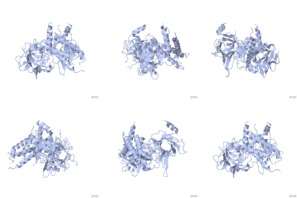
Ryanodine receptor
Ryanodine receptors (RyR) are the largest (over 5000 residues) ion channels and are responsible for the release of Ca+2 ions from the sarco/endoplasmic reticulum. RyR is a homotetramer which opens upon binding of Ca+2. RyR is modulated by various ions, small molecules and proteins. The protein modulators include calmodulin, FKBP, protein phosphatase and protin kinase A. The C-terminal of RyR forms the transmembrane ion-conducting pore. The N-terminal of RyR named “foot”, includes a trefoil knot fold and interacts with the various modulators. The corners of the RYR cytoplasmic area are called “clamp” and undergo conformational change upon opening or closing of the channel. RYR domains include three SPRY domains. SPRY2 forms the link between the N terminal gating ring and the clamp region. RYR1 and RYR2 have several phosphorylation sites. Ryanodine is a plant alkaloid which binds to RyR with high affinity and locks the channel in an open state. There are 3 RyR isoforms. RyR1 is found in skeletal muscles, RyR2 is found in cardiac muscles and RyR3 is found in the brain. FunctionDiseaseMutations in RyR are associated with skeletal muscle disorders and triggered cardiac arrhythmia. The N-terminal of RYR was found to be the disease hot spot of the molecule. RelevanceStructural highlights |
| ||||||||||
3D Structures of ryanodine receptor3D Structures of ryanodine receptor
Updated on 13-July-2015
