1dd3
CRYSTAL STRUCTURE OF RIBOSOMAL PROTEIN L12 FROM THERMOTOGA MARITIMACRYSTAL STRUCTURE OF RIBOSOMAL PROTEIN L12 FROM THERMOTOGA MARITIMA
Structural highlights
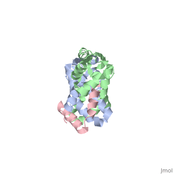
Evolutionary Conservation Check, as determined by ConSurfDB. You may read the explanation of the method and the full data available from ConSurf. Publication Abstract from PubMedProtein L12, the only multicopy component of the ribosome, is presumed to be involved in the binding of translation factors, stimulating factor-dependent GTP hydrolysis. Crystal structures of L12 from Thermotogamaritima have been solved in two space groups by the multiple anomalous dispersion method and refined at 2.4 and 2.0 A resolution. In both crystal forms, an asymmetric unit comprises two full-length L12 molecules and two N-terminal L12 fragments that are associated in a specific, hetero-tetrameric complex with one non-crystallographic 2-fold axis. The two full-length proteins form a tight, symmetric, parallel dimer, mainly through their N-terminal domains. Each monomer of this central dimer additionally associates in a different way with an N-terminal L12 fragment. Both dimerization modes are unlike models proposed previously and suggest that similar complexes may occur in vivo and in situ. The structures also display different L12 monomer conformations, in accord with the suggested dynamic role of the protein in the ribosomal translocation process. The structures have been submitted to the Protein Databank (http://www.rcsb.org/pdb) under accession numbers 1DD3 and 1DD4. Flexibility, conformational diversity and two dimerization modes in complexes of ribosomal protein L12.,Wahl MC, Bourenkov GP, Bartunik HD, Huber R EMBO J. 2000 Jan 17;19(2):174-86. PMID:10637222[1] From MEDLINE®/PubMed®, a database of the U.S. National Library of Medicine. See AlsoReferences
|
| ||||||||||||||