1qqw
| |||||||
| , resolution 2.75Å | |||||||
|---|---|---|---|---|---|---|---|
| Ligands: | |||||||
| Activity: | Catalase, with EC number 1.11.1.6 | ||||||
| Coordinates: | save as pdb, mmCIF, xml | ||||||
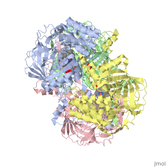
CRYSTAL STRUCTURE OF HUMAN ERYTHROCYTE CATALASE
OverviewOverview
Catalase (E.C. 1.11.1.6) was purified from human erythrocytes and crystallized in three different forms: orthorhombic, hexagonal and tetragonal. The structure of the orthorhombic crystal form of human erythrocyte catalase (HEC), with space group P2(1)2(1)2(1) and unit-cell parameters a = 84.9, b = 141.7, c = 232.5 A, was determined and refined with 2.75 A resolution data. Non-crystallographic symmetry restraints were employed and the resulting R value and R(free) were 0.206 and 0.272, respectively. The overall structure and arrangement of HEC molecules in the orthorhombic unit cell were very similar to those of bovine liver catalase (BLC). However, no NADPH was observed in the HEC crystal and a water was bound to the active-site residue His75. Conserved lattice interactions suggested a common growth mechanism for the orthorhombic crystals of HEC and BLC.
DiseaseDisease
Known disease associated with this structure: Acatalasemia OMIM:[115500]
About this StructureAbout this Structure
1QQW is a Single protein structure of sequence from Homo sapiens. The following page contains interesting information on the relation of 1QQW with [Catalase]. Full crystallographic information is available from OCA.
ReferenceReference
Structure of human erythrocyte catalase., Ko TP, Safo MK, Musayev FN, Di Salvo ML, Wang C, Wu SH, Abraham DJ, Acta Crystallogr D Biol Crystallogr. 2000 Feb;56(Pt 2):241-5. PMID:10666617
Page seeded by OCA on Thu Mar 20 13:41:28 2008