3r2a
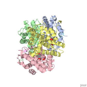
Crystal structure of RXRalpha ligand-binding domain complexed with corepressor SMRT2 and antagonist rheinCrystal structure of RXRalpha ligand-binding domain complexed with corepressor SMRT2 and antagonist rhein
Structural highlights
Publication Abstract from PubMedRetinoic X receptor (RXR) is a master nuclear receptor in the processes of cell development and homeostasis. Unliganded RXR exists in an auto-repressed tetramer, and agonists can induce RXR dimerization and coactivator recruitment for activation. However, the molecular mechanisms involving the corepressor recruitment and antagonist-mediated repression of RXR are still elusive. Here we reported the crystal structure of RXRalpha ligand-binding domain (LBD) complexed with silencing mediator for retinoid and thyroid hormone receptors (SMRT) corepressor motif. As the first structural report on the unliganded nuclear receptor bound to the corepressor motif, RXRalphaLBD-SMRT exhibited a significant structural rearrangement, compared with the apo RXRalphaLBD tetramer. To further elucidate the molecular determinant for RXR repression by antagonist, we also determined the crystal structure of RXRalphaLBD-SMRT complexed with the identified antagonist rhein. In the structure, two rhein molecules and two SMRT peptides were in the RXRalphaLBD tetramer, different from the case in RXRalphaLBD-SMRT structure, where four SMRT peptides bound to RXR tetramer. It seemed that rhein binding has resulted in a displacement of SMRT motif for activation function-2 (AF-2) motif binding to the receptor. Combining our current work with the published results, structural superposition of RXRalphaLBDs in different states revealed that RXR used an overlapped binding site for coactivator, corepressor and AF-2 motif, while AF-2 motif adopted different conformations for agonist or antagonist interaction, and coactivator or corepressor recruitment. Taken together, we thus proposed a molecular model of RXR repression on the tetramer. Structural basis for retinoic X receptor repression on the tetramer.,Zhang H, Chen L, Chen J, Jiang H, Shen X J Biol Chem. 2011 May 24. PMID:21613212[1] From MEDLINE®/PubMed®, a database of the U.S. National Library of Medicine. See AlsoReferences
|
| ||||||||||||||||||||