Protein Kinase A
Protein kinase A (PKA)Protein kinase A (PKA)
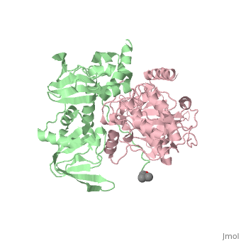
Protein kinase A (PKA) is a group of enzymes whose activity is dependent on the concentration of cAMP, and is also known as cAMP-dependent protein kinase. FunctionProtein kinase A is Protein Kinase A (PKA) helps animals cope with stressful situations. In the "flight or fight" response, muscles become ready for action as a result of the activity of PKA. Upon activation by cAMP, PKA alters the activities of target proteins by phopshorylating specific serine or threonine residues. This enzyme also helps regulate glucose levels. When activated by cAMP, PKA simultaneously catalyzes the breakdown of glycogen and inhibits the synthesis of glucose. PKA also helps regulate glycogen levels, sugar levels, and lipid metabolism. StructurePKA consists of two types of subunits. It has a 49-kd regulatory )(R) and a 38-kd (C).When PKA is enzymatically inactive (absence of cAMP), it is a tetrameric complex of a regulatory dimer bound to two catalytic subunits, forming the R2C2 complex. In its active state, the complex dissociates to form an R2 subunit and two C subunits. The catalytic subunit has two lobes. The larger lobe binds the peptide and contributes the key catalytic residues and the smaller lobe makes many contacts with ATP-Mg2+. MechanismPKA consists of a regulatory dimer, which each regulatory subunit attached to a catalytic subunit. When cAMP levels are low, the enzyme is intact, forming the R2C2 complex. However, when cAMP levels are high, two cAMP molecules bind to the regulatory sites of the complex, which leads to the dissociation of the R2C2 complex into an R2 subunit and two C subunits. When cAMP binds to the R chains, the binding allosterically removes the pseudostubstrate sequences, Arg-Arg-Gly-Ala-Ile, out of the catalytic sites. This psuedosubstrate sequence of R occupies the catalytic site of C and prevents it from interacting with protein substrates. The only difference between the pseudosubstrate sequence and the sequence for phophorylations is the Alanine in place of the serine. Once released, the two enzymatically active C subunites are free to phosphorylate proteins. DiseaseRelevanceStructural highlightsThis is a sample scene created with SAT to by Group, and another to make of the protein. You can make your own scenes on SAT starting from scratch or loading and editing one of these sample scenes.
|
| ||||||||||
ReferencesReferences
[1]Herraez A. Biomolecules in the computer: Jmol to the rescue. Biochem Mol Biol Educ. 2006 Jul;34(4):255-61. doi: 10.1002/bmb.2006.494034042644. PMID:21638687 doi:10.1002/bmb.2006.494034042644 [2]C. Kim, N.-H. Xuong, and S.S. Taylor, "Crystal structure of a complex between catalytic and regulatory (Rlα) subunits of PKA,"Science 307, 690 (2005) [doi:10.2210/rcsb_pdb/mom_2012_8]