Sandbox Reserved 814
RIBOSOMAL PROTEIN L14RIBOSOMAL PROTEIN L14
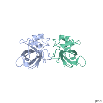
Introduction The L14 ribosomal protein is a protein which is included in the 50S subunit of the procaryote ribosome and in the 60s subunit of the eucaryote ribosome. The 50S and 60S subunits are the twos biggest subunits of the prokaryotic and eukaryotic ribosomes. They participate to the mechanism of translation by allowing the folding and stabilization of rRNA. The L14 subunit is one of the most conserved protein in the ribosome. L14 has been located at the 50S and 30S subunit interface between peptidyl transferase and GTPase regions. It allows contacts with the 16S rRNA of the 30S subunit (bridges B5 and B8) connecting the 2 subunits. StructureThe L14 subunit has a molecular mass of 13.3 kDa and contains 122 amino acids. It is composed of five stranded , a C-terminal region which contains two small and a beta-ribbon. The beta-barrel contains a hydrophobic core of highly conserved residues. The beta-barrel is stabilized by some hydrogen-bonding such as a bonding between loop 2 and loops 4 and 8. . Bidding sitesProtein-protein interactions are essential for the stability of ribosomes. The L14 subunits presents a perfect hydrophobic area on its structure too allow such an interaction with other ribosome’s subunits. This area is on the beta-barrel and is composed of : Leu25, Val40, Val57 and Ile2. This area is very exposed and separated from the RNA binding site. Links to L4, L7/L12, L10, L11, L17 and L19 has been showed and are allowed by this hydrophobic structure. In the L14 subunit, there are also two binding sites for the fixation of the rRNA. These two sites could each bind to a specific RNA sequence and induce the folding of the 23S rRNA. Role of the L14 subunitThe ribosome orchestrates the synthesis of proteins in all cells.The rRNA three dimensional organization is a major element in the activity of the ribonucleoprotein complex. This three dimensional structure is organized by the ribosomal proteins. Sequence alignment show that the structure of the L14 I highly conserved. It’s probably due to the fact that both mechanism and structure of the ribosome are common in all organisms.
By Bonhomme Clémence and Dilda Nathan |
| ||||||||||||||||
| This Sandbox is Reserved from 06/12/2018, through 30/06/2019 for use in the course "Structural Biology" taught by Bruno Kieffer at the University of Strasbourg, ESBS. This reservation includes Sandbox Reserved 1480 through Sandbox Reserved 1543. |
To get started:
More help: Help:Editing |