3hf1
| |||||||
| 3hf1, resolution 2.60Å () | |||||||
|---|---|---|---|---|---|---|---|
| Ligands: | , | ||||||
| Gene: | RRM2B, P53R2 (Homo sapiens) | ||||||
| Activity: | Ribonucleoside-diphosphate reductase, with EC number 1.17.4.1 | ||||||
| Related: | 1xsm, 2uw2, 1w69, 1w68 | ||||||
| |||||||
| |||||||
| Resources: | FirstGlance, OCA, RCSB, PDBsum | ||||||
| Coordinates: | save as pdb, mmCIF, xml | ||||||
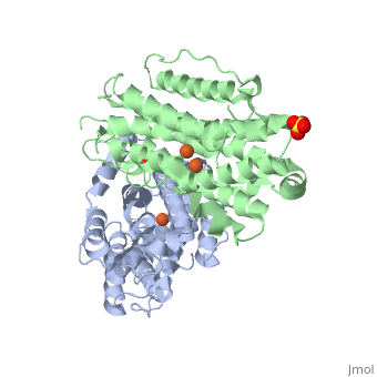

Crystal structure of human p53R2Crystal structure of human p53R2
DiseaseDisease
[RIR2B_HUMAN] Defects in RRM2B are the cause of mitochondrial DNA depletion syndrome type 8A (MTDPS8A) [MIM:612075]. A disorder due to mitochondrial dysfunction characterized by various combinations of neonatal hypotonia, neurological deterioration, respiratory distress, lactic acidosis, and renal tubulopathy.[1] [2] Defects in RRM2B are the cause of mitochondrial DNA depletion syndrome type 8B (MTDPS8B) [MIM:612075]. A disease due to mitochondrial dysfunction and characterized by ophthalmoplegia, ptosis, gastrointestinal dysmotility, cachexia, peripheral neuropathy. Defects in RRM2B are the cause of progressive external ophthalmoplegia with mitochondrial DNA deletions autosomal dominant type 5 (PEOA5) [MIM:613077]. A disorder characterized by progressive weakness of ocular muscles and levator muscle of the upper eyelid. In a minority of cases, it is associated with skeletal myopathy, which predominantly involves axial or proximal muscles and which causes abnormal fatigability and even permanent muscle weakness. Ragged-red fibers and atrophy are found on muscle biopsy. A large proportion of chronic ophthalmoplegias are associated with other symptoms, leading to a multisystemic pattern of this disease. Additional symptoms are variable, and may include cataracts, hearing loss, sensory axonal neuropathy, ataxia, depression, hypogonadism, and parkinsonism.[3]
FunctionFunction
[RIR2B_HUMAN] Plays a pivotal role in cell survival by repairing damaged DNA in a p53/TP53-dependent manner. Supplies deoxyribonucleotides for DNA repair in cells arrested at G1 or G2. Contains an iron-tyrosyl free radical center required for catalysis. Forms an active ribonucleotide reductase (RNR) complex with RRM1 which is expressed both in resting and proliferating cells in response to DNA damage.[4] [5] [6]
About this StructureAbout this Structure
3hf1 is a 2 chain structure with sequence from Homo sapiens. Full crystallographic information is available from OCA.
ReferenceReference
- ↑ Smith P, Zhou B, Ho N, Yuan YC, Su L, Tsai SC, Yen Y. 2.6 X-ray Crystal Structure of Human p53R2. Biochemistry. 2009 Sep 3. PMID:19728742 doi:10.1021/bi9001425
- ↑ Bourdon A, Minai L, Serre V, Jais JP, Sarzi E, Aubert S, Chretien D, de Lonlay P, Paquis-Flucklinger V, Arakawa H, Nakamura Y, Munnich A, Rotig A. Mutation of RRM2B, encoding p53-controlled ribonucleotide reductase (p53R2), causes severe mitochondrial DNA depletion. Nat Genet. 2007 Jun;39(6):776-80. Epub 2007 May 7. PMID:17486094 doi:10.1038/ng2040
- ↑ Bornstein B, Area E, Flanigan KM, Ganesh J, Jayakar P, Swoboda KJ, Coku J, Naini A, Shanske S, Tanji K, Hirano M, DiMauro S. Mitochondrial DNA depletion syndrome due to mutations in the RRM2B gene. Neuromuscul Disord. 2008 Jun;18(6):453-9. doi: 10.1016/j.nmd.2008.04.006. Epub, 2008 May 27. PMID:18504129 doi:10.1016/j.nmd.2008.04.006
- ↑ Tyynismaa H, Ylikallio E, Patel M, Molnar MJ, Haller RG, Suomalainen A. A heterozygous truncating mutation in RRM2B causes autosomal-dominant progressive external ophthalmoplegia with multiple mtDNA deletions. Am J Hum Genet. 2009 Aug;85(2):290-5. doi: 10.1016/j.ajhg.2009.07.009. Epub 2009 , Aug 6. PMID:19664747 doi:10.1016/j.ajhg.2009.07.009
- ↑ Tanaka H, Arakawa H, Yamaguchi T, Shiraishi K, Fukuda S, Matsui K, Takei Y, Nakamura Y. A ribonucleotide reductase gene involved in a p53-dependent cell-cycle checkpoint for DNA damage. Nature. 2000 Mar 2;404(6773):42-9. PMID:10716435 doi:10.1038/35003506
- ↑ Guittet O, Hakansson P, Voevodskaya N, Fridd S, Graslund A, Arakawa H, Nakamura Y, Thelander L. Mammalian p53R2 protein forms an active ribonucleotide reductase in vitro with the R1 protein, which is expressed both in resting cells in response to DNA damage and in proliferating cells. J Biol Chem. 2001 Nov 2;276(44):40647-51. Epub 2001 Aug 21. PMID:11517226 doi:10.1074/jbc.M106088200
- ↑ Yamaguchi T, Matsuda K, Sagiya Y, Iwadate M, Fujino MA, Nakamura Y, Arakawa H. p53R2-dependent pathway for DNA synthesis in a p53-regulated cell cycle checkpoint. Cancer Res. 2001 Nov 15;61(22):8256-62. PMID:11719458