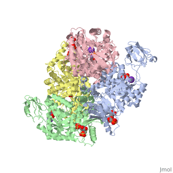
2vgb
| |||||||||
| 2vgb, resolution 2.73Å () | |||||||||
|---|---|---|---|---|---|---|---|---|---|
| Ligands: | , , , | ||||||||
| Activity: | Pyruvate kinase, with EC number 2.7.1.40 | ||||||||
| Related: | 1liy, 1liu, 1liw, 1lix | ||||||||
| |||||||||
| |||||||||
| Resources: | FirstGlance, OCA, RCSB, PDBsum | ||||||||
| Coordinates: | save as pdb, mmCIF, xml | ||||||||

HUMAN ERYTHROCYTE PYRUVATE KINASEHUMAN ERYTHROCYTE PYRUVATE KINASE
Template:ABSTRACT PUBMED 11960989
About this StructureAbout this Structure
2vgb is a 4 chain structure with sequence from Homo sapiens. This structure supersedes the now removed PDB entry 1liu. Full crystallographic information is available from OCA.
See AlsoSee Also
ReferenceReference
- ↑ Valentini G, Chiarelli LR, Fortin R, Dolzan M, Galizzi A, Abraham DJ, Wang C, Bianchi P, Zanella A, Mattevi A. Structure and function of human erythrocyte pyruvate kinase. Molecular basis of nonspherocytic hemolytic anemia. J Biol Chem. 2002 Jun 28;277(26):23807-14. Epub 2002 Apr 17. PMID:11960989 doi:10.1074/jbc.M202107200