File:Imatinib bound to receptor.PNG
Imatinib_bound_to_receptor.PNG (529 × 221 pixels, file size: 9 KB, MIME type: image/png)
LicensingLicensing
{{subst:No license from license selector|Don't know}}
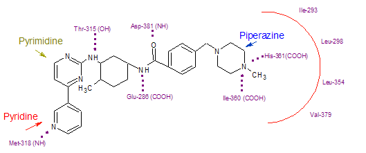
Imatinib binds to Abl domain via six hydrogen bond interactions. This stabilizes the imatinib Bcr-Abl complex and prevents ATP from reaching its binding site.[3][7][9] The hydrogen bonds involve the pyridine-N and backbone-NH of Met-318, the aminopyrimidine and side chain hydroxyl of Thr-315, the amide-NH and side chain carboxylate of Glu-285, the carbonyl and backbone-NH of Asp-381, the protonated methylpiperazine with the backbone-carbonyl atoms of Ile-360 and His-361. Additionally, a number of van der Waals interactions contribute to binding.[7] A hydrophobic pocket is formed by amino acid residues Ile-293, Leu-298, Leu-354 and Val-379 around the phenyl ring adjacent to the piperazinyl-methyl group of imatinib.[9] At the time of its discovery, in the absence of structural information, no clear explanation for the impressive selectivity of imatinib could be found
File history
Click on a date/time to view the file as it appeared at that time.
| Date/Time | Thumbnail | Dimensions | User | Comment | |
|---|---|---|---|---|---|
| current | 21:24, 9 December 2012 | | 529 × 221 (9 KB) | Cristina Murga (talk | contribs) |
You cannot overwrite this file.
File usage
The following page uses this file: