1qav
|
UNEXPECTED MODES OF PDZ DOMAIN SCAFFOLDING REVEALED BY STRUCTURE OF NNOS-SYNTROPHIN COMPLEX
OverviewOverview
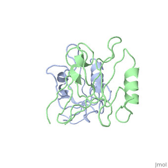
The PDZ protein interaction domain of neuronal nitric oxide synthase (nNOS) can heterodimerize with the PDZ domains of postsynaptic density protein 95 and syntrophin through interactions that are not mediated by recognition of a typical carboxyl-terminal motif. The nNOS-syntrophin PDZ complex structure revealed that the domains interact in an unusual linear head-to-tail arrangement. The nNOS PDZ domain has two opposite interaction surfaces-one face has the canonical peptide binding groove, whereas the other has a beta-hairpin "finger." This nNOS beta finger docks in the syntrophin peptide binding groove, mimicking a peptide ligand, except that a sharp beta turn replaces the normally required carboxyl terminus. This structure explains how PDZ domains can participate in diverse interaction modes to assemble protein networks.
About this StructureAbout this Structure
1QAV is a Protein complex structure of sequences from Mus musculus and Rattus norvegicus. Full crystallographic information is available from OCA.
ReferenceReference
Unexpected modes of PDZ domain scaffolding revealed by structure of nNOS-syntrophin complex., Hillier BJ, Christopherson KS, Prehoda KE, Bredt DS, Lim WA, Science. 1999 Apr 30;284(5415):812-5. PMID:10221915
Page seeded by OCA on Thu Feb 21 14:37:41 2008