1atg
|
AZOTOBACTER VINELANDII PERIPLASMIC MOLYBDATE-BINDING PROTEIN
OverviewOverview
Background:. Periplasmic receptors constitute a diverse class of binding, proteins that differ widely in size, sequence and ligand specificity., Nevertheless, almost all of them display a common beta/alpha folding motif, and have similar tertiary structures consisting of two globular domains., The ligand is bound at the bottom of a deep cleft, which lies at the, interface between these two domains. The oxyanion-binding proteins are, notable in that they can discriminate between very similar ligands., Results:. Azotobacter vinelandii is unusual in that it possesses two, periplasmic molybdate-binding proteins. The crystal structure of one of, these with bound ligand has been determined at 1.2 A resolution. It, superficially resembles the structure of sulphate-binding protein (SBP), from ... [(full description)]
About this StructureAbout this Structure
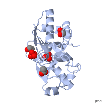
1ATG is a [Single protein] structure of sequence from [Azotobacter vinelandii] with WO4, ACT, SO4 and EDO as [ligands]. Structure known Active Site: MOB. Full crystallographic information is available from [OCA].
ReferenceReference
Ligand size is a major determinant of specificity in periplasmic oxyanion-binding proteins: the 1.2 A resolution crystal structure of Azotobacter vinelandii ModA., Lawson DM, Williams CE, Mitchenall LA, Pau RN, Structure. 1998 Dec 15;6(12):1529-39. PMID:9862806
Page seeded by OCA on Tue Oct 30 12:59:14 2007