Sandbox SouthUniversity1
|
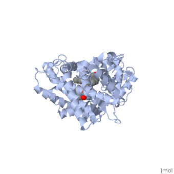
This tutorial covers the basic structure and function of the Cytochrome P450 (CYP) enzymes, the most important group of drug metabolizing enzymes. The example used here for illustration purposes is the second protein discovered in the CYP 1 family, in subfamily A (generally referred to as CYP1A2). This protein is shown in an interactive window below.
Turn off (toggle) spinning of the protein by clicking on the button below the structure. The quality of the molecule rendering can also be increased by clicking the "toggle quality" button, although this may decrease the smoothness when the molecule is rotating.
Now rotate the molecule by clicking and dragging in the window with your cursor. Resize the molecule by holding down the shift key and dragging up and down. Rotate and resize the molecule until you can clearly see that there are 2 molecules shown in a "space-filling" representation in the middle of the protein. These are a heme molecule, which is absolutely vital for the enzyme's function, and a second molecule (alpha-naphthoflavone) which represents a drug that is about to be metabolized.
First, lets look at the heme. Click the following link to hide the rest of the protein and Carbon atoms are shown in grey, Nitrogen atoms are blue, Oxygen is red, and the central iron atom is orange. The iron atom is the vital center that does the actual oxidation of substrates (drugs or other xenobiotics). It is highlighted
Now look at the substrate molecule being oxidized and view its orientation relative to the heme group by clicking Resize and rotate the molecules until you can see how the two molecules are oriented in relationship to each other.
The flavone is metabolized (oxidized) by introduction of a hydroxy group onto the phenyl ring that is attached to the tricyclic ring system.
References The structure of CYP1A2 shown above is found in the Research Collaboration of Structural Bioinformatics Protein Database as entry 2HI4.