Malate dehydrogenase
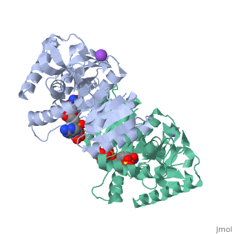
Malate Dehydrogenase (MDH)(PDB entry 2x0i) is most known for its role in the metabolic pathway in the tricarboxylic acid cycle or Kreb's Cycle which is critical to cellular respiration in cells [1]; however, the enzyme is also in many other metabolic pathways such as glyoxylate bypass, amino acid synthesis, glucogenesis, and oxidation/reduction balance [1]. It is classified as a oxidoreductase[2]. Malate Dehydrogenase has been extensively studied due to its many isozymes [2]. The enzyme exists in two places inside a cell, in the mitochondria and cytoplasm. In the mitochondria, the enzyme catalyzes the reaction of malate to oxaloacetate; but in the cytoplasm, the enzyme catalyzes oxaloacetate to malate to allow transport [3]. This conversion is an essential catalytic step in each different metabolic mechanism. The enzyme malate dehydrogenase is composed of either a dimer or tetramer depending on the location and organism [4]. During catalysis, the enzyme subunits are non-cooperative between active sites. The mitochondrial MDH is complexly, allosterically controlled by citrate, but no other known metabolic regulation mechanisms have been discovered. Further, the exact mechanism of regulation has yet to be discovered [5]. Kinetically, the pH of optimization is 7.6 for oxaloacetate conversion and 9.6 for malate conversion. The reported K(m) value for malate conversion is 215 uM and the V(max) value is 87.8 uM/min [6].
| |||||||
| 2x0i, resolution 2.91Å () | |||||||
|---|---|---|---|---|---|---|---|
| Ligands: | , , | ||||||
| Activity: | Malate dehydrogenase, with EC number 1.1.1.37 | ||||||
| Related: | 2x06, 2x0j, 2x0n, 2x0r, 2x0s | ||||||
| |||||||
| |||||||
| Resources: | FirstGlance, OCA, RCSB, PDBsum | ||||||
| Coordinates: | save as pdb, mmCIF, xml | ||||||

StructureStructure
The secondary structure of a single subunit contains a wrapped by . Near the sodium bound end, 4 small anti-parallel beta sheets and 1 small alpha helix enable a turn in the residue chain. Opposite the sodium bound ligand, 6 alpha helices point towards a common point, three on each side of the beta sheet backbone. The alpha helices form a for a NAD+ cofactor to attach near the beta sheeting. The structure most nearly resembles an alternating alpha/beta classification. As for the 3D structure, the enzyme forms a sort of for the substrate to bind.
MechanismMechanism
The mechanism of catalysis is dependent on . These residues are HIS 195 and ASP 168 which are involved in hydrogen bonding, ASP 53 associated with , and a triad of arginine residues at 102, 109, and 171. During the conversation of malate to oxaloacetate, a key conformational change occurs on the binding of substrate in which a “loop” flips into an up position to block the active site from the solvent.
 When this occurs, the other residues in the active site are brought closer to the substrate to enable the conversion. R102 and R109 are involved in this loop flip and thus invariant. After the loop flip, the malate complex is stabilized via hydrogen bonding before accepting a proton transfer from NADH to form oxaloacetate [7].
Evolutionary DivergenceEvolutionary Divergence
The evolutionary past of MDH shows a divergence to form lactate dehydrogenase (LDH) which functions in a very similar way to MDH. Although there is a very low sequence conservation among MDH and LDH’s [3] the structure of the enzyme has remained relatively conserved. The key difference between the two is in the substrate: LDH catalyzes pyruvate to lactate.
3D Structures of Malate Dehydrogenase3D Structures of Malate Dehydrogenase
The holo-MDH contains NAD or its derivatives while the apo-MDH lacks it.
Holo-MDHHolo-MDH
2x0r – HmMDH (mutant)+NAD - Haloarcula marismortui 1o6z - HmMDH (mutant)+NADH 1hlp – HmMDH+NAD 1x0i – AfMDH+NADH – Archaeoglobus fulgidus 2x0j - AfMDH+etheno-NAD 1hlp – HmMDH+NAD 1x0i – AfMDH+NADH 2x0j - AfMDH+etheno-NAD 1ib6, 1ie3 – EcMDH (mutant)+NAD - Escherichia coli 1eMDH – EcMDH+NAD+citrate 1cMDH - EcMDH+citrate 3i0p – MDH+NAD – Entamoeba histolytica 3gvh – BmMDH+NAD – Brucella melitensis 3gvi - BmMDH+ADP 2hjr – MDH+adenosine diphosphoribose – Cryptosporidium parvum 2dfd – MDH+NAD – human type 2 1wze – TfMDH (mutant)+NAD – Thermus flavus 1wzi - TfMDH (mutant)+NDP 1bdm - TfMDH (mutant)+beta-6-hydroxy-1,4,5,6-tetrhydronicotinamide adenine dinucleotide 1bMDH – TfMDH+NAD 1y7t – TtMDH+NADPH – Thermus thermophilus 2cvq - TtMDH+NADP 1v9n – MDH+NADPH – Pyrococcus horikoshii 1z2i – MDH+NAD – Agrobacterium tumefaciens 1uxg, 1uxh, 1uxi, 1uxj, 1uxk, 1ur5 – MDH (mutant)+NAD – Chloroflexus aurantiacus 1guz, 1guy, 1gv0 – CvMDH+NAD – Chlorobium vibrioforme 1civ – MDH+NADP – Flaveria bidentis 1b8u, 1b8v – AaMDH+NAD - Aquaspirillum arcticum 5MDHh – SsMDH+NAD+alpha-ketomalonic acid – Sus scrofa 4MDHh – SsMDH+NAD
apo-MDHapo-MDH
2j5r, 2j5k, 2j5q, 1d3a – HmMDH 2hlp – HmMDH (mutant) 3hhp, 2pwz – EcMDH 3fi9 – MDH – Porphyromonas gingivalis 3d5t - MDH – Burkholderia pseudomallei 2d4a – MDH – Aeropyrum pernix 1iz9 - TtMDH 1sev, 1smk – MDH – Citrullus lanatus 1gv1 – CvMDH 1b8p – AaMDH 7MDHh – MDH – Sorgum bicolor 1mld – SsMDH
Additional ResourcesAdditional Resources
For additional information, see: Carbohydrate Metabolism
ReferencesReferences
- ↑ Minarik P, Tomaskova N, Kollarova M, Antalik M. Malate dehydrogenases--structure and function. Gen Physiol Biophys. 2002 Sep;21(3):257-65. PMID:12537350
- ↑ Matsuda T, Takahashi-Yanaga F, Yoshihara T, Maenaka K, Watanabe Y, Miwa Y, Morimoto S, Kubohara Y, Hirata M, Sasaguri T. Dictyostelium Differentiation-Inducing Factor-1 Binds to Mitochondrial Malate Dehydrogenase and Inhibits Its Activity. J Pharmacol Sci. 2010 Feb 20. PMID:20173310
- ↑ Matsuda T, Takahashi-Yanaga F, Yoshihara T, Maenaka K, Watanabe Y, Miwa Y, Morimoto S, Kubohara Y, Hirata M, Sasaguri T. Dictyostelium Differentiation-Inducing Factor-1 Binds to Mitochondrial Malate Dehydrogenase and Inhibits Its Activity. J Pharmacol Sci. 2010 Feb 20. PMID:20173310
- ↑ Musrati RA, Kollarova M, Mernik N, Mikulasova D. Malate dehydrogenase: distribution, function and properties. Gen Physiol Biophys. 1998 Sep;17(3):193-210. PMID:9834842
- ↑ Boernke WE, Millard CS, Stevens PW, Kakar SN, Stevens FJ, Donnelly MI. Stringency of substrate specificity of Escherichia coli malate dehydrogenase. Arch Biochem Biophys. 1995 Sep 10;322(1):43-52. PMID:7574693 doi:http://dx.doi.org/10.1006/abbi.1995.1434
- ↑ Plancarte A, Nava G, Mendoza-Hernandez G. Purification, properties, and kinetic studies of cytoplasmic malate dehydrogenase from Taenia solium cysticerci. Parasitol Res. 2009 Jul;105(1):175-83. Epub 2009 Mar 10. PMID:19277715 doi:10.1007/s00436-009-1380-6
- ↑ Goward CR, Nicholls DJ. Malate dehydrogenase: a model for structure, evolution, and catalysis. Protein Sci. 1994 Oct;3(10):1883-8. PMID:7849603 doi:http://dx.doi.org/10.1002/pro.5560031027