User:Wayne Decatur/3fpn Morph methods
Moving to match Figure 3Moving to match Figure 3
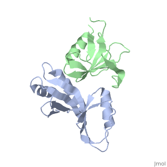
Using Pymol and the 3fpn file, I moved so interface is perpendicular to y axis:
translate [10,0,0], chain b
rotate y, 65, chain b
Saved molecule.
Morph from normal 3fpn structure to view in Figure 3 of article describing the structureMorph from normal 3fpn structure to view in Figure 3 of article describing the structure
|
Took the two files and submitted them. Since the structures didn't have nucleic acids, I took the advice here and used the Yale Morph Server for morphing complexes.
Uploaded to Proteopedia File:3fpntorotatedversion.pdb.
loaded '3fpntorotatedversion.pdb' in Scene Authoring Tools.
[Note: Control the animation with the 'animation' submenu on the menu that comes up if you click on the Jmol frank in the bottom rigth corner. ]
Paper on the structurePaper on the structure
- ↑ Pakotiprapha D, Liu Y, Verdine GL, Jeruzalmi D. A structural model for the damage-sensing complex in bacterial nucleotide excision repair. J Biol Chem. 2009 May 8;284(19):12837-44. Epub 2009 Mar 13. PMID:19287003 doi:10.1074/jbc.M900571200