M2 Proton Channel
M2 Proton Channel from Influenza A VirusM2 Proton Channel from Influenza A Virus
|
BackgroundBackground
The M2 proton channel is a key protein that leads to viral infection.[1] The M2 proton channel acidifies the viron which allows the viral matrix protein (M1) to disassociate from the ribonucleoprotein (RNP).[2] This allows the RNP to be transported to the nucleus of the cell.[2] Several recent studies have looked at the effects of [3] and [4] on inhibiting the transfer of protons through the M2 channel.[3] It has been found that M2 is resistant to these two drugs in 90% of humans, birds and pigs.[3] Understanding the structure and function of this proton channel is necessary in solving the resistance problem.[3] The of the M2 proton channel influenza A virus was solved in 2008 (PBD: 3bkd).[3]
StructureStructure
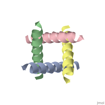
The M2 proton channel from influenza A is 97 amino acid residues and forms a 24-residue N-terminal extracellular domain, a 19-residue trans-membrane domain, and a 54-residue C-terminal cytoplasmic domain.[2] The 19-residue TM domain forms the highly selective proton channel.[1] Circular dichroism spectra has shown the TM domain to form an that spans the membrane.[2] By analytical ultracentrifugation, the TM domain is found to form which contains four identical α-helices.[1] Secondary structure is color coded by Alpha Helices. When viewed in the the N-terminus is located near the extracellular side of the membrane while the C-terminus is located near the cytosolic side of the membrane. This tetrameric bundle of the TM domain is found by NMR to be tilted by 25-38° from the channel axis.[1] The trameric helices form a left-handed bundle that resembles a truncated cone.[3] The TM helicies are arranged around the channel pore with an approximate four-fold rotational symmetry.[1]
Central CavityCentral Cavity
|
The hydrophilic residues in each α-helix monomer are oriented towards the pore lumen.[2] The residues will be in contact with the membrane (Color code= Hydrophobic). Most of the residues in the M2 channel are hydrophobic except Ser31 Gly34, and His37.[2] The central cavity of the M2 channel is a water-filled pore that is interrupted at residue in each monomer.[5] Mutagenesis studies have found that the residues facing the pore are Val27, Ala30, Ser31, Gly34, His37, Leu38, and Trp41.[2] Residues His37 and play a key role in the gating mechanism and the selectivity filter.[2] In the , the His37 and Trp41 residues block the channel, preventing proton conductance.
pH GatingpH Gating
The M2 channel is low-pH gated and has a 50-fold increase in proton conductance when the pH drops from 8.2 down to 4.2.[2] The His side chain, a five-membered ring, has two nitrogen atoms.[1] At a pH of 7 or higher, only one nitrogen is protonated in the neutral imidazole form.[1] However, at a pH of about 5.8, the second nitrogen becomes protonated and forms the cationic imidazolium form.[1] This protonation causes the His residues in each monomer to move away from each other due to electrostatic repulsion.[1][5][2] This allows the channel to open and allow water and protons to cross the membrane.[5][2]
SelectivitySelectivity
The selectivity of this channel is based on the binding of a proton to the His37 residues in each of the α-helices in the tetramer.[1][5][2] Without protons binding to the His residues, the channel would not open. Also, because there is an excess amount of protons when the pH is in acidic conditions on the extracellular side of the membrane, a proton gradient is formed. Once the channel is open, the protons move with their gradient to the cytosolic side of the membrane. These protons are then used to acidify the viron.[2]
ReferencesReferences
- ↑ 1.00 1.01 1.02 1.03 1.04 1.05 1.06 1.07 1.08 1.09 Takeuchi H, Okada A, Miura T. Roles of the histidine and tryptophan side chains in the M2 proton channel from influenza A virus. FEBS Lett. 2003 Sep 18;552(1):35-8. PMID:12972149
- ↑ 2.00 2.01 2.02 2.03 2.04 2.05 2.06 2.07 2.08 2.09 2.10 2.11 2.12 Wu Y, Voth GA. Computational studies of proton transport through the M2 channel. FEBS Lett. 2003 Sep 18;552(1):23-7. PMID:12972147
- ↑ 3.0 3.1 3.2 3.3 3.4 3.5 Stouffer AL, Acharya R, Salom D, Levine AS, Di Costanzo L, Soto CS, Tereshko V, Nanda V, Stayrook S, DeGrado WF. Structural basis for the function and inhibition of an influenza virus proton channel. Nature. 2008 Jan 31;451(7178):596-9. PMID:18235504 doi:10.1038/nature06528
- ↑ Schnell JR, Chou JJ. Structure and mechanism of the M2 proton channel of influenza A virus. Nature. 2008 Jan 31;451(7178):591-5. PMID:18235503 doi:10.1038/nature06531
- ↑ 5.0 5.1 5.2 5.3 Lear JD. Proton conduction through the M2 protein of the influenza A virus; a quantitative, mechanistic analysis of experimental data. FEBS Lett. 2003 Sep 18;552(1):17-22. PMID:12972146