M2 Proton Channel from Influenza A VirusM2 Proton Channel from Influenza A Virus
BackgroundBackground
The M2 proton channel is a key protein that leads to viral infection [Takeuchi et al]. The M2 proton channel acidifies the viron which allows the viral matrix protein (M1) to disassociate from the ribonucleoprotein (RNP).[1] This allows the RNP to be transported to the nucleus of the cell.[1] Several recent studies have looked at the effects of [2] and [3] on inhibiting the transfer of protons through the M2 channel.[2] It has been found that M2 is resistant to these two drugs in 90% of humans, birds and pigs.[2] Understanding the structure and function of this proton channel is necessary in solving the resistance problem.[2]
StructureStructure
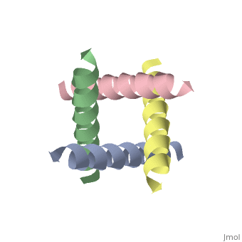
The M2 proton channel from influenza A is 97 amino acid residues and forms a 24-residue N-terminal extracellular domain, a 19-residue trans-membrane domain, and a 54-residue C-terminal cytoplasmic domain [wu et al]. The 19-residue TM domain forms the highly selective proton channel.[4] Circular dichroism spectra has shown the TM domain to form one α-helix that spans the membrane.[1] By analytical ultracentrifugation, the TM domain is found to form [4] This tetrameric bundle of the TM domain is found by NMR to be tilted by 25-38° from the channel axis.[4] The trameric helices form a left-handed bundle that resembles a truncated cone.[2] The TM helicies are arranged around the channel pore with an approximate fourfold rotational symmetry.[4]
Central CavityCentral Cavity
pH GatingpH Gating
ReferencesReferences
- ↑ 1.0 1.1 1.2 Wu Y, Voth GA. Computational studies of proton transport through the M2 channel. FEBS Lett. 2003 Sep 18;552(1):23-7. PMID:12972147
- ↑ 2.0 2.1 2.2 2.3 2.4 Stouffer AL, Acharya R, Salom D, Levine AS, Di Costanzo L, Soto CS, Tereshko V, Nanda V, Stayrook S, DeGrado WF. Structural basis for the function and inhibition of an influenza virus proton channel. Nature. 2008 Jan 31;451(7178):596-9. PMID:18235504 doi:10.1038/nature06528
- ↑ Schnell JR, Chou JJ. Structure and mechanism of the M2 proton channel of influenza A virus. Nature. 2008 Jan 31;451(7178):591-5. PMID:18235503 doi:10.1038/nature06531
- ↑ 4.0 4.1 4.2 4.3 Takeuchi H, Okada A, Miura T. Roles of the histidine and tryptophan side chains in the M2 proton channel from influenza A virus. FEBS Lett. 2003 Sep 18;552(1):35-8. PMID:12972149
proteopedia link