1yv0
Crystal structure of skeletal muscle troponin in the Ca2+-free stateCrystal structure of skeletal muscle troponin in the Ca2+-free state
Structural highlights
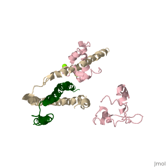
Function[TNNT3_CHICK] Troponin T is the tropomyosin-binding subunit of troponin, the thin filament regulatory complex which confers calcium-sensitivity to striated muscle actomyosin ATPase activity. [TNNC2_CHICK] Troponin is the central regulatory protein of striated muscle contraction. Tn consists of three components: Tn-I which is the inhibitor of actomyosin ATPase, Tn-T which contains the binding site for tropomyosin and Tn-C. The binding of calcium to Tn-C abolishes the inhibitory action of Tn on actin filaments. [TNNI2_CHICK] Troponin I is the inhibitory subunit of troponin, the thin filament regulatory complex which confers calcium-sensitivity to striated muscle actomyosin ATPase activity. Evolutionary Conservation Check, as determined by ConSurfDB. You may read the explanation of the method and the full data available from ConSurf. Publication Abstract from PubMedTroponin senses Ca2+ to regulate contraction in striated muscle. Structures of skeletal muscle troponin composed of TnC (the sensor), TnI (the regulator), and TnT (the link to the muscle thin filament) have been determined. The structure of troponin in the Ca(2+)-activated state features a nearly twofold symmetrical assembly of TnI and TnT subunits penetrated asymmetrically by the dumbbell-shaped TnC subunit. Ca ions are thought to regulate contraction by controlling the presentation to and withdrawal of the TnI inhibitory segment from the thin filament. Here, we show that the rigid central helix of the sensor binds the inhibitory segment of TnI in the Ca(2+)-activated state. Comparison of crystal structures of troponin in the Ca(2+)-activated state at 3.0 angstroms resolution and in the Ca(2+)-free state at 7.0 angstroms resolution shows that the long framework helices of TnI and TnT, presumed to be a Ca(2+)-independent structural domain of troponin are unchanged. Loss of Ca ions causes the rigid central helix of the sensor to collapse and to release the inhibitory segment of TnI. The inhibitory segment of TnI changes conformation from an extended loop in the presence of Ca2+ to a short alpha-helix in its absence. We also show that Anapoe, a detergent molecule, increases the contractile force of muscle fibers and binds specifically, together with the TnI switch helix, in a hydrophobic pocket of TnC upon activation by Ca ions. Ca(2+)-regulated structural changes in troponin.,Vinogradova MV, Stone DB, Malanina GG, Karatzaferi C, Cooke R, Mendelson RA, Fletterick RJ Proc Natl Acad Sci U S A. 2005 Apr 5;102(14):5038-43. Epub 2005 Mar 22. PMID:15784741[1] From MEDLINE®/PubMed®, a database of the U.S. National Library of Medicine. See AlsoReferences |
| ||||||||||||||||||||