1c05
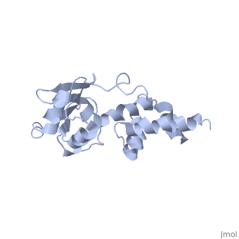
SOLUTION STRUCTURE OF RIBOSOMAL PROTEIN S4 DELTA 41, REFINED WITH DIPOLAR COUPLINGS (MINIMIZED AVERAGE STRUCTURE)SOLUTION STRUCTURE OF RIBOSOMAL PROTEIN S4 DELTA 41, REFINED WITH DIPOLAR COUPLINGS (MINIMIZED AVERAGE STRUCTURE)
Structural highlights
Function[RS4_GEOSE] One of the primary rRNA binding proteins, it binds directly to 16S rRNA where it nucleates assembly of the body of the 30S subunit (By similarity).[HAMAP-Rule:MF_01306_B] With S5 and S12 plays an important role in translational accuracy (By similarity).[HAMAP-Rule:MF_01306_B] Evolutionary Conservation Check, as determined by ConSurfDB. You may read the explanation of the method and the full data available from ConSurf. Publication Abstract from PubMedProkaryotic protein S4 initiates assembly of the small ribosomal subunit by binding to 16 S rRNA. Residues 43-200 of S4 from Bacillus stearothermophilus (S4 Delta41) bind to both 16 S rRNA and to a mRNA pseudoknot. In order to obtain structure-based insights regarding RNA binding, we previously determined the solution structure of S4 Delta41 using NOE, hydrogen bond, and torsion angle restraints. S4 Delta41 is elongated, with two distinct subdomains, one all helical, the other including a beta-sheet. In contrast to the high resolution structures obtained for each individual subdomain, their relative orientation was not precisely defined because only 17 intersubdomain NOE restraints were determined. Compared to the 1.7 A crystal structure, when the sheet-containing subdomains are superimposed, the helical subdomain is twisted by almost 45 degrees about the long axis of the molecule in the solution structure. Because variations in subdomain orientation may explain how the protein recognizes multiple RNA targets, our current goal is to determine the orientation of the subdomains in solution with high precision. To this end, NOE assignments were re-examined. NOESY experiments on a specifically labeled sample revealed that one of the intersubdomain restraints had been misassigned. However, the revised set of NOE restraints produces solution structures that still have imprecisely defined subdomain orientations and that lie between the original NMR structure and the crystal structure. In contrast, augmenting the NOE restraints with N-H dipolar couplings, measured in uniaxial liquid crystalline phases, clearly establishes the relative orientation of the subdomains. Data obtained from two independent liquid crystalline milieux, DMPC/DHPC bicelles and the filamentous bacteriophage Pf1, show that the relative orientation of the subdomains in solution is quite similar to the subdomain orientation in the crystal structure. The solution structure, refined with dipolar data, is presented and its implications for S4's RNA binding activity are discussed. Refining the overall structure and subdomain orientation of ribosomal protein S4 delta41 with dipolar couplings measured by NMR in uniaxial liquid crystalline phases.,Markus MA, Gerstner RB, Draper DE, Torchia DA J Mol Biol. 1999 Sep 17;292(2):375-87. PMID:10493882[1] From MEDLINE®/PubMed®, a database of the U.S. National Library of Medicine. See AlsoReferences
|
| ||||||||||||||||