5dfr
| |||||||
| , resolution 2.3Å | |||||||
|---|---|---|---|---|---|---|---|
| Ligands: | |||||||
| Activity: | Dihydrofolate reductase, with EC number 1.5.1.3 | ||||||
| Coordinates: | save as pdb, mmCIF, xml | ||||||
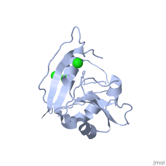
CRYSTAL STRUCTURE OF UNLIGANDED ESCHERICHIA COLI DIHYDROFOLATE REDUCTASE. LIGAND-INDUCED CONFORMATIONAL CHANGES AND COOPERATIVITY IN BINDING
OverviewOverview
The crystal structure of unliganded dihydrofolate reductase (DHFR) from Escherichia coli has been solved and refined to an R factor of 19% at 2.3-A resolution in a crystal form that is nonisomorphous with each of the previously reported E. coli DHFR crystal structures [Bolin, J. T., Filman, D. J., Matthews, D. A., Hamlin, B. C., & Kraut, J. (1982) J. Biol. Chem. 257, 13650-13662; Bystroff, C., Oatley, S. J., & Kraut, J. (1990) Biochemistry 29, 3263-3277]. Significant conformational changes occur between the apoenzyme and each of the complexes: the NADP+ holoenzyme, the folate-NADP+ ternary complex, and the methotrexate (MTX) binary complex. The changes are small, with the largest about 3 A and most of them less than 1 A. For simplicity a two-domain description is adopted in which one domain contains the NADP+ 2'-phosphate binding site and the binding sites for the rest of the coenzyme and for the substrate lie between the two domains. Binding of either NADP+ or MTX induces a closing of the PABG-binding cleft and realignment of alpha-helices C and F which bind the pyrophosphate of the coenzyme. Formation of the ternary complex from the holoenzyme does not involve further relative domain shifts but does involve a shift of alpha-helix B and a floppy loop (the Met-20 loop) that precedes alpha B. These observations suggest a mechanism for cooperativity in binding between substrate and coenzyme wherein the greatest degree of cooperativity is expressed in the transition-state complex. We explore the idea that the MTX binary complex in some ways resembles the transition-state complex.
About this StructureAbout this Structure
5DFR is a Single protein structure of sequence from Escherichia coli. Full crystallographic information is available from OCA.
ReferenceReference
Crystal structure of unliganded Escherichia coli dihydrofolate reductase. Ligand-induced conformational changes and cooperativity in binding., Bystroff C, Kraut J, Biochemistry. 1991 Feb 26;30(8):2227-39. PMID:1998681
Page seeded by OCA on Thu Mar 20 19:11:53 2008