Rebecca Martin/Sandbox1: Difference between revisions
Jump to navigation
Jump to search
No edit summary |
No edit summary |
||
| Line 124: | Line 124: | ||
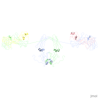
<applet load='1r70' size='300' frame='true' align='right' caption='monomeric IgA2' /> <applet load='1iga' size='300' frame='true' align='right' caption='monomeric IgA1' /> | <applet load='1r70' size='300' frame='true' align='right' caption='monomeric IgA2' /> <applet load='1iga' size='300' frame='true' align='right' caption='monomeric IgA1' /> | ||
Revision as of 23:40, 22 April 2009
IgAIgA
IgA1
StructureStructure
- Immunoglobulin Structure
Antibodies are composed of a heavy chain and a light chain.
Fab fragment
- Forms of IgA
Dimeric Structure
|
- Secretory Component
IgA1 and IgA2IgA1 and IgA2
|
|
|
|
Secretory ComponentSecretory Component
|
Insights into FunctionInsights into Function
EvolutionEvolution
Implications in Science and MedicineImplications in Science and Medicine
Limitations of the Current StudiesLimitations of the Current Studies
- Because of the nature of the IgA molecule, crystalizing this structure was not possible. Therefore, many of these structures are based on models and not actual crystal structures. Because ...., the models were depositable in the PDB. I tried to include other crystallographic data when available, supporting the proposed models- as the authors did in the original papers.
Questions for the FutureQuestions for the Future
- Because of the limitating resolution of these models, many details concerning the binding residues and residue interactions are left unknown. Crystallographic structure will yield further insights into the structure of IgA, the interactions between IgA and other molecules, and ....
LinksLinks
IgAIgA
- Monomeric
- Fab and Fc Fragments
- Refined crystal structure of the galactan-binding immunoglobulin fab j539 at 1.95-angstroms resolution 2fbj
- Phosphocholine binding immunoglobulin fab mc/pc603. an x-ray diffraction study at 2.7 angstroms 1mcp
- Phosphocholine binding immunoglobulin fab mc/pc603. an x-ray diffraction study at 3.1 angstroms 2mcp
- Crystal structure of human FcaRI bound to IgA1-Fc 1ow0
- Refined crystal structure of a recombinant immunoglobulin domain and a complementarity-determining region 1-grafted mutant 2imm and2imn
- Dimeric and Secretory
ReceptorsReceptors
- Crystal Structure of a Ligand-Binding Domain of the Human Polymeric Ig Receptor, pIgR 1XED
- Crystal structure of human FcaRI 10vz
- Crystal structure of a Staphylococcus aureus protein (SSL7) in complex with Fc of human IgA1 2qej
Other Isotypes (for comparison)Other Isotypes (for comparison)
- IgM: Solution structure of human Immunoglobulin M 2rcj
- IgG:
- IgD:
- IgE: