2dfx: Difference between revisions
Jump to navigation
Jump to search
No edit summary |
No edit summary |
||
| Line 1: | Line 1: | ||
[[Image:2dfx. | {{Seed}} | ||
[[Image:2dfx.png|left|200px]] | |||
<!-- | <!-- | ||
| Line 9: | Line 10: | ||
{{STRUCTURE_2dfx| PDB=2dfx | SCENE= }} | {{STRUCTURE_2dfx| PDB=2dfx | SCENE= }} | ||
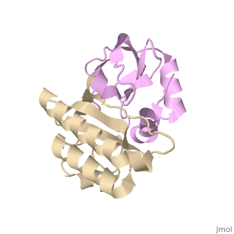
===Crystal structure of the carboxy terminal domain of colicin E5 complexed with its inhibitor=== | |||
<!-- | |||
The line below this paragraph, {{ABSTRACT_PUBMED_17099236}}, adds the Publication Abstract to the page | |||
(as it appears on PubMed at http://www.pubmed.gov), where 17099236 is the PubMed ID number. | |||
--> | |||
{{ABSTRACT_PUBMED_17099236}} | |||
==About this Structure== | ==About this Structure== | ||
| Line 30: | Line 34: | ||
[[Category: Alpha/beta protein]] | [[Category: Alpha/beta protein]] | ||
[[Category: Protein-inhibitor protein complex]] | [[Category: Protein-inhibitor protein complex]] | ||
''Page seeded by [http://oca.weizmann.ac.il/oca OCA ] on | |||
''Page seeded by [http://oca.weizmann.ac.il/oca OCA ] on Mon Jul 28 09:29:52 2008'' | |||
Revision as of 09:29, 28 July 2008
| |||||||||
| 2dfx, resolution 1.90Å () | |||||||||
|---|---|---|---|---|---|---|---|---|---|
| Related: | 2djh | ||||||||
| |||||||||
| |||||||||
| |||||||||
| Resources: | FirstGlance, OCA, PDBsum, RCSB | ||||||||
| Coordinates: | save as pdb, mmCIF, xml | ||||||||

Crystal structure of the carboxy terminal domain of colicin E5 complexed with its inhibitorCrystal structure of the carboxy terminal domain of colicin E5 complexed with its inhibitor
Template:ABSTRACT PUBMED 17099236
About this StructureAbout this Structure
2DFX is a Protein complex structure of sequences from Escherichia coli. Full crystallographic information is available from OCA.
ReferenceReference
Structural basis for sequence-dependent recognition of colicin E5 tRNase by mimicking the mRNA-tRNA interaction., Yajima S, Inoue S, Ogawa T, Nonaka T, Ohsawa K, Masaki H, Nucleic Acids Res. 2006;34(21):6074-82. Epub 2006 Nov 11. PMID:17099236
Page seeded by OCA on Mon Jul 28 09:29:52 2008