2hbs: Difference between revisions
No edit summary |
No edit summary |
||
| Line 3: | Line 3: | ||
<StructureSection load='2hbs' size='340' side='right'caption='[[2hbs]], [[Resolution|resolution]] 2.05Å' scene=''> | <StructureSection load='2hbs' size='340' side='right'caption='[[2hbs]], [[Resolution|resolution]] 2.05Å' scene=''> | ||
== Structural highlights == | == Structural highlights == | ||
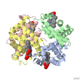
<table><tr><td colspan='2'>[[2hbs]] is a 8 chain structure with sequence from [ | <table><tr><td colspan='2'>[[2hbs]] is a 8 chain structure with sequence from [https://en.wikipedia.org/wiki/Homo_sapiens Homo sapiens]. The May 2003 RCSB PDB [https://pdb.rcsb.org/pdb/static.do?p=education_discussion/molecule_of_the_month/index.html Molecule of the Month] feature on ''Hemoglobin'' by Shuchismita Dutta and David S. Goodsell is [https://dx.doi.org/10.2210/rcsb_pdb/mom_2003_5 10.2210/rcsb_pdb/mom_2003_5]. Full crystallographic information is available from [http://oca.weizmann.ac.il/oca-bin/ocashort?id=2HBS OCA]. For a <b>guided tour on the structure components</b> use [https://proteopedia.org/fgij/fg.htm?mol=2HBS FirstGlance]. <br> | ||
</td></tr><tr id='ligand'><td class="sblockLbl"><b>[[Ligand|Ligands:]]</b></td><td class="sblockDat" id="ligandDat"><scene name='pdbligand=HEM:PROTOPORPHYRIN+IX+CONTAINING+FE'>HEM</scene></td></tr> | </td></tr><tr id='method'><td class="sblockLbl"><b>[[Empirical_models|Method:]]</b></td><td class="sblockDat" id="methodDat">X-ray diffraction, [[Resolution|Resolution]] 2.05Å</td></tr> | ||
<tr id='resources'><td class="sblockLbl"><b>Resources:</b></td><td class="sblockDat"><span class='plainlinks'>[ | <tr id='ligand'><td class="sblockLbl"><b>[[Ligand|Ligands:]]</b></td><td class="sblockDat" id="ligandDat"><scene name='pdbligand=HEM:PROTOPORPHYRIN+IX+CONTAINING+FE'>HEM</scene></td></tr> | ||
<tr id='resources'><td class="sblockLbl"><b>Resources:</b></td><td class="sblockDat"><span class='plainlinks'>[https://proteopedia.org/fgij/fg.htm?mol=2hbs FirstGlance], [http://oca.weizmann.ac.il/oca-bin/ocaids?id=2hbs OCA], [https://pdbe.org/2hbs PDBe], [https://www.rcsb.org/pdb/explore.do?structureId=2hbs RCSB], [https://www.ebi.ac.uk/pdbsum/2hbs PDBsum], [https://prosat.h-its.org/prosat/prosatexe?pdbcode=2hbs ProSAT]</span></td></tr> | |||
</table> | </table> | ||
== Disease == | == Disease == | ||
[ | [https://www.uniprot.org/uniprot/HBA_HUMAN HBA_HUMAN] Defects in HBA1 may be a cause of Heinz body anemias (HEIBAN) [MIM:[https://omim.org/entry/140700 140700]. This is a form of non-spherocytic hemolytic anemia of Dacie type 1. After splenectomy, which has little benefit, basophilic inclusions called Heinz bodies are demonstrable in the erythrocytes. Before splenectomy, diffuse or punctate basophilia may be evident. Most of these cases are probably instances of hemoglobinopathy. The hemoglobin demonstrates heat lability. Heinz bodies are observed also with the Ivemark syndrome (asplenia with cardiovascular anomalies) and with glutathione peroxidase deficiency.<ref>PMID:2833478</ref> Defects in HBA1 are the cause of alpha-thalassemia (A-THAL) [MIM:[https://omim.org/entry/604131 604131]. The thalassemias are the most common monogenic diseases and occur mostly in Mediterranean and Southeast Asian populations. The hallmark of alpha-thalassemia is an imbalance in globin-chain production in the adult HbA molecule. The level of alpha chain production can range from none to very nearly normal levels. Deletion of both copies of each of the two alpha-globin genes causes alpha(0)-thalassemia, also known as homozygous alpha thalassemia. Due to the complete absence of alpha chains, the predominant fetal hemoglobin is a tetramer of gamma-chains (Bart hemoglobin) that has essentially no oxygen carrying capacity. This causes oxygen starvation in the fetal tissues leading to prenatal lethality or early neonatal death. The loss of three alpha genes results in high levels of a tetramer of four beta chains (hemoglobin H), causing a severe and life-threatening anemia known as hemoglobin H disease. Untreated, most patients die in childhood or early adolescence. The loss of two alpha genes results in mild alpha-thalassemia, also known as heterozygous alpha-thalassemia. Affected individuals have small red cells and a mild anemia (microcytosis). If three of the four alpha-globin genes are functional, individuals are completely asymptomatic. Some rare forms of alpha-thalassemia are due to point mutations (non-deletional alpha-thalassemia). The thalassemic phenotype is due to unstable globin alpha chains that are rapidly catabolized prior to formation of the alpha-beta heterotetramers. Note=Alpha(0)-thalassemia is associated with non-immune hydrops fetalis, a generalized edema of the fetus with fluid accumulation in the body cavities due to non-immune causes. Non-immune hydrops fetalis is not a diagnosis in itself but a symptom, a feature of many genetic disorders, and the end-stage of a wide variety of disorders. Defects in HBA1 are the cause of hemoglobin H disease (HBH) [MIM:[https://omim.org/entry/613978 613978]. HBH is a form of alpha-thalassemia due to the loss of three alpha genes. This results in high levels of a tetramer of four beta chains (hemoglobin H), causing a severe and life-threatening anemia. Untreated, most patients die in childhood or early adolescence.<ref>PMID:10569720</ref> | ||
== Function == | == Function == | ||
[ | [https://www.uniprot.org/uniprot/HBA_HUMAN HBA_HUMAN] Involved in oxygen transport from the lung to the various peripheral tissues. | ||
== Evolutionary Conservation == | == Evolutionary Conservation == | ||
[[Image:Consurf_key_small.gif|200px|right]] | [[Image:Consurf_key_small.gif|200px|right]] | ||
| Line 21: | Line 22: | ||
</jmol>, as determined by [http://consurfdb.tau.ac.il/ ConSurfDB]. You may read the [[Conservation%2C_Evolutionary|explanation]] of the method and the full data available from [http://bental.tau.ac.il/new_ConSurfDB/main_output.php?pdb_ID=2hbs ConSurf]. | </jmol>, as determined by [http://consurfdb.tau.ac.il/ ConSurfDB]. You may read the [[Conservation%2C_Evolutionary|explanation]] of the method and the full data available from [http://bental.tau.ac.il/new_ConSurfDB/main_output.php?pdb_ID=2hbs ConSurf]. | ||
<div style="clear:both"></div> | <div style="clear:both"></div> | ||
==See Also== | ==See Also== | ||
*[[Hemoglobin|Hemoglobin]] | *[[Hemoglobin|Hemoglobin]] | ||
*[[Hemoglobin 3D structures|Hemoglobin 3D structures]] | *[[Hemoglobin 3D structures|Hemoglobin 3D structures]] | ||
*[[Sandbox 12345|Sandbox 12345]] | *[[Sandbox 12345|Sandbox 12345]] | ||
== References == | == References == | ||
<references/> | <references/> | ||
| Line 45: | Line 35: | ||
[[Category: Large Structures]] | [[Category: Large Structures]] | ||
[[Category: RCSB PDB Molecule of the Month]] | [[Category: RCSB PDB Molecule of the Month]] | ||
[[Category: Adachi | [[Category: Adachi K]] | ||
[[Category: Harrington | [[Category: Harrington DJ]] | ||
[[Category: Junior | [[Category: Royer Junior WE]] | ||
Latest revision as of 12:31, 14 February 2024
THE HIGH RESOLUTION CRYSTAL STRUCTURE OF DEOXYHEMOGLOBIN STHE HIGH RESOLUTION CRYSTAL STRUCTURE OF DEOXYHEMOGLOBIN S
Structural highlights
DiseaseHBA_HUMAN Defects in HBA1 may be a cause of Heinz body anemias (HEIBAN) [MIM:140700. This is a form of non-spherocytic hemolytic anemia of Dacie type 1. After splenectomy, which has little benefit, basophilic inclusions called Heinz bodies are demonstrable in the erythrocytes. Before splenectomy, diffuse or punctate basophilia may be evident. Most of these cases are probably instances of hemoglobinopathy. The hemoglobin demonstrates heat lability. Heinz bodies are observed also with the Ivemark syndrome (asplenia with cardiovascular anomalies) and with glutathione peroxidase deficiency.[1] Defects in HBA1 are the cause of alpha-thalassemia (A-THAL) [MIM:604131. The thalassemias are the most common monogenic diseases and occur mostly in Mediterranean and Southeast Asian populations. The hallmark of alpha-thalassemia is an imbalance in globin-chain production in the adult HbA molecule. The level of alpha chain production can range from none to very nearly normal levels. Deletion of both copies of each of the two alpha-globin genes causes alpha(0)-thalassemia, also known as homozygous alpha thalassemia. Due to the complete absence of alpha chains, the predominant fetal hemoglobin is a tetramer of gamma-chains (Bart hemoglobin) that has essentially no oxygen carrying capacity. This causes oxygen starvation in the fetal tissues leading to prenatal lethality or early neonatal death. The loss of three alpha genes results in high levels of a tetramer of four beta chains (hemoglobin H), causing a severe and life-threatening anemia known as hemoglobin H disease. Untreated, most patients die in childhood or early adolescence. The loss of two alpha genes results in mild alpha-thalassemia, also known as heterozygous alpha-thalassemia. Affected individuals have small red cells and a mild anemia (microcytosis). If three of the four alpha-globin genes are functional, individuals are completely asymptomatic. Some rare forms of alpha-thalassemia are due to point mutations (non-deletional alpha-thalassemia). The thalassemic phenotype is due to unstable globin alpha chains that are rapidly catabolized prior to formation of the alpha-beta heterotetramers. Note=Alpha(0)-thalassemia is associated with non-immune hydrops fetalis, a generalized edema of the fetus with fluid accumulation in the body cavities due to non-immune causes. Non-immune hydrops fetalis is not a diagnosis in itself but a symptom, a feature of many genetic disorders, and the end-stage of a wide variety of disorders. Defects in HBA1 are the cause of hemoglobin H disease (HBH) [MIM:613978. HBH is a form of alpha-thalassemia due to the loss of three alpha genes. This results in high levels of a tetramer of four beta chains (hemoglobin H), causing a severe and life-threatening anemia. Untreated, most patients die in childhood or early adolescence.[2] FunctionHBA_HUMAN Involved in oxygen transport from the lung to the various peripheral tissues. Evolutionary Conservation Check, as determined by ConSurfDB. You may read the explanation of the method and the full data available from ConSurf. See AlsoReferences
|
| ||||||||||||||||||