2x0n: Difference between revisions
No edit summary |
No edit summary |
||
| Line 3: | Line 3: | ||
<StructureSection load='2x0n' size='340' side='right'caption='[[2x0n]], [[Resolution|resolution]] 3.20Å' scene=''> | <StructureSection load='2x0n' size='340' side='right'caption='[[2x0n]], [[Resolution|resolution]] 3.20Å' scene=''> | ||
== Structural highlights == | == Structural highlights == | ||
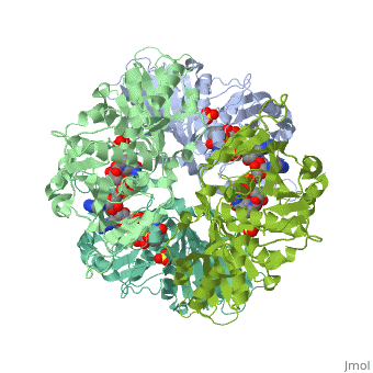
<table><tr><td colspan='2'>[[2x0n]] is a 6 chain structure with sequence from [ | <table><tr><td colspan='2'>[[2x0n]] is a 6 chain structure with sequence from [https://en.wikipedia.org/wiki/Trypanosoma_brucei_brucei Trypanosoma brucei brucei]. This structure supersedes the now removed PDB entry [http://oca.weizmann.ac.il/oca-bin/send-pdb?obs=1&id=1gga 1gga]. Full crystallographic information is available from [http://oca.weizmann.ac.il/oca-bin/ocashort?id=2X0N OCA]. For a <b>guided tour on the structure components</b> use [https://proteopedia.org/fgij/fg.htm?mol=2X0N FirstGlance]. <br> | ||
</td></tr><tr id=' | </td></tr><tr id='method'><td class="sblockLbl"><b>[[Empirical_models|Method:]]</b></td><td class="sblockDat" id="methodDat">X-ray diffraction, [[Resolution|Resolution]] 3.2Å</td></tr> | ||
<tr id=' | <tr id='ligand'><td class="sblockLbl"><b>[[Ligand|Ligands:]]</b></td><td class="sblockDat" id="ligandDat"><scene name='pdbligand=NAD:NICOTINAMIDE-ADENINE-DINUCLEOTIDE'>NAD</scene>, <scene name='pdbligand=SO4:SULFATE+ION'>SO4</scene></td></tr> | ||
<tr id='resources'><td class="sblockLbl"><b>Resources:</b></td><td class="sblockDat"><span class='plainlinks'>[ | <tr id='resources'><td class="sblockLbl"><b>Resources:</b></td><td class="sblockDat"><span class='plainlinks'>[https://proteopedia.org/fgij/fg.htm?mol=2x0n FirstGlance], [http://oca.weizmann.ac.il/oca-bin/ocaids?id=2x0n OCA], [https://pdbe.org/2x0n PDBe], [https://www.rcsb.org/pdb/explore.do?structureId=2x0n RCSB], [https://www.ebi.ac.uk/pdbsum/2x0n PDBsum], [https://prosat.h-its.org/prosat/prosatexe?pdbcode=2x0n ProSAT]</span></td></tr> | ||
</table> | </table> | ||
== Function == | |||
[https://www.uniprot.org/uniprot/G3PG_TRYBB G3PG_TRYBB] | |||
== Evolutionary Conservation == | == Evolutionary Conservation == | ||
[[Image:Consurf_key_small.gif|200px|right]] | [[Image:Consurf_key_small.gif|200px|right]] | ||
| Line 36: | Line 38: | ||
[[Category: Large Structures]] | [[Category: Large Structures]] | ||
[[Category: Trypanosoma brucei brucei]] | [[Category: Trypanosoma brucei brucei]] | ||
[[Category: Hajdu | [[Category: Hajdu J]] | ||
[[Category: Hol | [[Category: Hol WGJ]] | ||
[[Category: Vellieux | [[Category: Vellieux FMD]] | ||
Latest revision as of 13:21, 20 December 2023
Structure of glycosomal glyceraldehyde-3-phosphate dehydrogenase from Trypanosoma brucei determined from Laue dataStructure of glycosomal glyceraldehyde-3-phosphate dehydrogenase from Trypanosoma brucei determined from Laue data
Structural highlights
FunctionEvolutionary Conservation Check, as determined by ConSurfDB. You may read the explanation of the method and the full data available from ConSurf. Publication Abstract from PubMedThe three-dimensional structure of glycosomal glyceraldehyde-3-phosphate dehydrogenase [D-glyceraldehyde-3-phosphate:NAD+ oxidoreductase (phosphorylating), EC 1.12.1.12] from the sleeping-sickness parasite Trypanosoma brucei was solved by molecular replacement at 3.2-A resolution with an x-ray data set collected by the Laue method. For data collection, three crystals were exposed to the polychromatic synchrotron x-ray beam for a total of 20.5 sec. The structure was solved by using the Bacillus stearothermophilus enzyme model [Skarzynski, T., Moody, P. C. E. & Wonacott, A. J. (1987) J. Mol. Biol. 193, 171-187] with a partial data set which was 37% complete. The crystals contain six subunits per asymmetric unit, which allowed us to overcome the absence of > 60% of the reflections by 6-fold density averaging. After molecular dynamics refinement, the current molecular model has an R factor of 17.6%. Comparing the structure of the trypanosome enzyme with that of the homologous human muscle enzyme, which was determined at 2.4-A resolution, reveals important structural differences in the NAD binding region. These are of great interest for the design of specific inhibitors of the parasite enzyme. Structure of glycosomal glyceraldehyde-3-phosphate dehydrogenase from Trypanosoma brucei determined from Laue data.,Vellieux FM, Hajdu J, Verlinde CL, Groendijk H, Read RJ, Greenhough TJ, Campbell JW, Kalk KH, Littlechild JA, Watson HC, et al. Proc Natl Acad Sci U S A. 1993 Mar 15;90(6):2355-9. PMID:8460146[1] From MEDLINE®/PubMed®, a database of the U.S. National Library of Medicine. See AlsoReferences
|
| ||||||||||||||||||