2gox: Difference between revisions
No edit summary |
No edit summary |
||
| Line 3: | Line 3: | ||
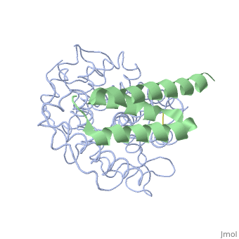
<StructureSection load='2gox' size='340' side='right'caption='[[2gox]], [[Resolution|resolution]] 2.20Å' scene=''> | <StructureSection load='2gox' size='340' side='right'caption='[[2gox]], [[Resolution|resolution]] 2.20Å' scene=''> | ||
== Structural highlights == | == Structural highlights == | ||
<table><tr><td colspan='2'>[[2gox]] is a 4 chain structure with sequence from [https://en.wikipedia.org/wiki/ | <table><tr><td colspan='2'>[[2gox]] is a 4 chain structure with sequence from [https://en.wikipedia.org/wiki/Homo_sapiens Homo sapiens] and [https://en.wikipedia.org/wiki/Staphylococcus_aureus_subsp._aureus_Mu50 Staphylococcus aureus subsp. aureus Mu50]. Full crystallographic information is available from [http://oca.weizmann.ac.il/oca-bin/ocashort?id=2GOX OCA]. For a <b>guided tour on the structure components</b> use [https://proteopedia.org/fgij/fg.htm?mol=2GOX FirstGlance]. <br> | ||
</td></tr><tr id=' | </td></tr><tr id='method'><td class="sblockLbl"><b>[[Empirical_models|Method:]]</b></td><td class="sblockDat" id="methodDat">X-ray diffraction, [[Resolution|Resolution]] 2.2Å</td></tr> | ||
<tr id='resources'><td class="sblockLbl"><b>Resources:</b></td><td class="sblockDat"><span class='plainlinks'>[https://proteopedia.org/fgij/fg.htm?mol=2gox FirstGlance], [http://oca.weizmann.ac.il/oca-bin/ocaids?id=2gox OCA], [https://pdbe.org/2gox PDBe], [https://www.rcsb.org/pdb/explore.do?structureId=2gox RCSB], [https://www.ebi.ac.uk/pdbsum/2gox PDBsum], [https://prosat.h-its.org/prosat/prosatexe?pdbcode=2gox ProSAT]</span></td></tr> | <tr id='resources'><td class="sblockLbl"><b>Resources:</b></td><td class="sblockDat"><span class='plainlinks'>[https://proteopedia.org/fgij/fg.htm?mol=2gox FirstGlance], [http://oca.weizmann.ac.il/oca-bin/ocaids?id=2gox OCA], [https://pdbe.org/2gox PDBe], [https://www.rcsb.org/pdb/explore.do?structureId=2gox RCSB], [https://www.ebi.ac.uk/pdbsum/2gox PDBsum], [https://prosat.h-its.org/prosat/prosatexe?pdbcode=2gox ProSAT]</span></td></tr> | ||
</table> | </table> | ||
== Disease == | == Disease == | ||
[https://www.uniprot.org/uniprot/CO3_HUMAN CO3_HUMAN] Defects in C3 are the cause of complement component 3 deficiency (C3D) [MIM:[https://omim.org/entry/613779 613779]. A rare defect of the complement classical pathway. Patients develop recurrent, severe, pyogenic infections because of ineffective opsonization of pathogens. Some patients may also develop autoimmune disorders, such as arthralgia and vasculitic rashes, lupus-like syndrome and membranoproliferative glomerulonephritis.<ref>PMID:19913840</ref> <ref>PMID:9596584</ref> <ref>PMID:11387479</ref> <ref>PMID:15713468</ref> <ref>PMID:7961791</ref> [:] Genetic variation in C3 is associated with susceptibility to age-related macular degeneration type 9 (ARMD9) [MIM:[https://omim.org/entry/611378 611378]. ARMD is a multifactorial eye disease and the most common cause of irreversible vision loss in the developed world. In most patients, the disease is manifest as ophthalmoscopically visible yellowish accumulations of protein and lipid that lie beneath the retinal pigment epithelium and within an elastin-containing structure known as Bruch membrane.<ref>PMID:19913840</ref> <ref>PMID:17634448</ref> Defects in C3 are a cause of susceptibility to hemolytic uremic syndrome atypical type 5 (AHUS5) [MIM:[https://omim.org/entry/612925 612925]. An atypical form of hemolytic uremic syndrome. It is a complex genetic disease characterized by microangiopathic hemolytic anemia, thrombocytopenia, renal failure and absence of episodes of enterocolitis and diarrhea. In contrast to typical hemolytic uremic syndrome, atypical forms have a poorer prognosis, with higher death rates and frequent progression to end-stage renal disease. Note=Susceptibility to the development of atypical hemolytic uremic syndrome can be conferred by mutations in various components of or regulatory factors in the complement cascade system. Other genes may play a role in modifying the phenotype.<ref>PMID:19913840</ref> <ref>PMID:18796626</ref> <ref>PMID:20513133</ref> Note=Increased levels of C3 and its cleavage product ASP, are associated with obesity, diabetes and coronary heart disease. Short-term endurance training reduces baseline ASP levels and subsequently fat storage.<ref>PMID:19913840</ref> | |||
== Function == | == Function == | ||
[https://www.uniprot.org/uniprot/CO3_HUMAN CO3_HUMAN] C3 plays a central role in the activation of the complement system. Its processing by C3 convertase is the central reaction in both classical and alternative complement pathways. After activation C3b can bind covalently, via its reactive thioester, to cell surface carbohydrates or immune aggregates.<ref>PMID:8376604</ref> <ref>PMID:2909530</ref> <ref>PMID:9059512</ref> <ref>PMID:9555951</ref> <ref>PMID:10432298</ref> <ref>PMID:15833747</ref> <ref>PMID:16333141</ref> <ref>PMID:19615750</ref> Derived from proteolytic degradation of complement C3, C3a anaphylatoxin is a mediator of local inflammatory process. It induces the contraction of smooth muscle, increases vascular permeability and causes histamine release from mast cells and basophilic leukocytes.<ref>PMID:8376604</ref> <ref>PMID:2909530</ref> <ref>PMID:9059512</ref> <ref>PMID:9555951</ref> <ref>PMID:10432298</ref> <ref>PMID:15833747</ref> <ref>PMID:16333141</ref> <ref>PMID:19615750</ref> Acylation stimulating protein (ASP): adipogenic hormone that stimulates triglyceride (TG) synthesis and glucose transport in adipocytes, regulating fat storage and playing a role in postprandial TG clearance. Appears to stimulate TG synthesis via activation of the PLC, MAPK and AKT signaling pathways. Ligand for GPR77. Promotes the phosphorylation, ARRB2-mediated internalization and recycling of GPR77.<ref>PMID:8376604</ref> <ref>PMID:2909530</ref> <ref>PMID:9059512</ref> <ref>PMID:9555951</ref> <ref>PMID:10432298</ref> <ref>PMID:15833747</ref> <ref>PMID:16333141</ref> <ref>PMID:19615750</ref> | |||
== Evolutionary Conservation == | == Evolutionary Conservation == | ||
[[Image:Consurf_key_small.gif|200px|right]] | [[Image:Consurf_key_small.gif|200px|right]] | ||
| Line 39: | Line 38: | ||
__TOC__ | __TOC__ | ||
</StructureSection> | </StructureSection> | ||
[[Category: | [[Category: Homo sapiens]] | ||
[[Category: Large Structures]] | [[Category: Large Structures]] | ||
[[Category: | [[Category: Staphylococcus aureus subsp. aureus Mu50]] | ||
[[Category: Geisbrecht | [[Category: Geisbrecht BV]] | ||
[[Category: Hammel | [[Category: Hammel M]] | ||
Revision as of 12:45, 30 August 2023
Crystal structure of Efb-C / C3d ComplexCrystal structure of Efb-C / C3d Complex
Structural highlights
DiseaseCO3_HUMAN Defects in C3 are the cause of complement component 3 deficiency (C3D) [MIM:613779. A rare defect of the complement classical pathway. Patients develop recurrent, severe, pyogenic infections because of ineffective opsonization of pathogens. Some patients may also develop autoimmune disorders, such as arthralgia and vasculitic rashes, lupus-like syndrome and membranoproliferative glomerulonephritis.[1] [2] [3] [4] [5] [:] Genetic variation in C3 is associated with susceptibility to age-related macular degeneration type 9 (ARMD9) [MIM:611378. ARMD is a multifactorial eye disease and the most common cause of irreversible vision loss in the developed world. In most patients, the disease is manifest as ophthalmoscopically visible yellowish accumulations of protein and lipid that lie beneath the retinal pigment epithelium and within an elastin-containing structure known as Bruch membrane.[6] [7] Defects in C3 are a cause of susceptibility to hemolytic uremic syndrome atypical type 5 (AHUS5) [MIM:612925. An atypical form of hemolytic uremic syndrome. It is a complex genetic disease characterized by microangiopathic hemolytic anemia, thrombocytopenia, renal failure and absence of episodes of enterocolitis and diarrhea. In contrast to typical hemolytic uremic syndrome, atypical forms have a poorer prognosis, with higher death rates and frequent progression to end-stage renal disease. Note=Susceptibility to the development of atypical hemolytic uremic syndrome can be conferred by mutations in various components of or regulatory factors in the complement cascade system. Other genes may play a role in modifying the phenotype.[8] [9] [10] Note=Increased levels of C3 and its cleavage product ASP, are associated with obesity, diabetes and coronary heart disease. Short-term endurance training reduces baseline ASP levels and subsequently fat storage.[11] FunctionCO3_HUMAN C3 plays a central role in the activation of the complement system. Its processing by C3 convertase is the central reaction in both classical and alternative complement pathways. After activation C3b can bind covalently, via its reactive thioester, to cell surface carbohydrates or immune aggregates.[12] [13] [14] [15] [16] [17] [18] [19] Derived from proteolytic degradation of complement C3, C3a anaphylatoxin is a mediator of local inflammatory process. It induces the contraction of smooth muscle, increases vascular permeability and causes histamine release from mast cells and basophilic leukocytes.[20] [21] [22] [23] [24] [25] [26] [27] Acylation stimulating protein (ASP): adipogenic hormone that stimulates triglyceride (TG) synthesis and glucose transport in adipocytes, regulating fat storage and playing a role in postprandial TG clearance. Appears to stimulate TG synthesis via activation of the PLC, MAPK and AKT signaling pathways. Ligand for GPR77. Promotes the phosphorylation, ARRB2-mediated internalization and recycling of GPR77.[28] [29] [30] [31] [32] [33] [34] [35] Evolutionary Conservation Check, as determined by ConSurfDB. You may read the explanation of the method and the full data available from ConSurf. Publication Abstract from PubMedTo provide insight into bacterial suppression of complement-mediated immunity, we present here structures of a bacterial complement inhibitory protein, both free and bound to its complement target. The 1.25-A structure of the complement component C3-inhibitory domain of Staphylococcus aureus extracellular fibrinogen-binding protein (Efb-C) demonstrated a helical motif involved in complement regulation, whereas the 2.2-A structure of Efb-C bound to the C3d domain of human C3 allowed insight into the recognition of complement proteins by invading pathogens. Our structure-function studies provided evidence for a previously unrecognized mode of complement inhibition whereby Efb-C binds to native C3 and alters the solution conformation of C3 in a way that renders it unable to participate in successful 'downstream' activation of the complement response. A structural basis for complement inhibition by Staphylococcus aureus.,Hammel M, Sfyroera G, Ricklin D, Magotti P, Lambris JD, Geisbrecht BV Nat Immunol. 2007 Apr;8(4):430-7. Epub 2007 Mar 11. PMID:17351618[36] From MEDLINE®/PubMed®, a database of the U.S. National Library of Medicine. See AlsoReferences
|
| ||||||||||||||||