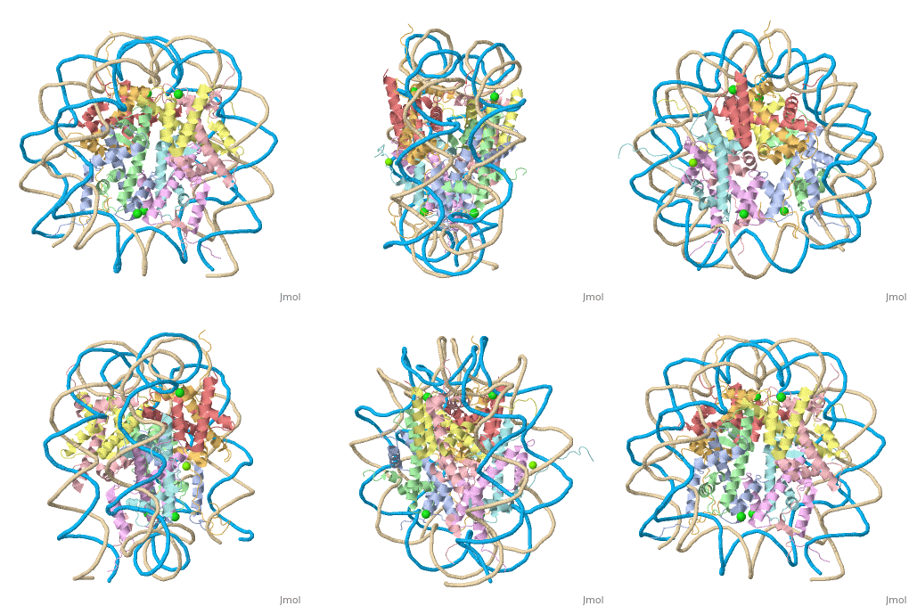
5f99: Difference between revisions
No edit summary |
No edit summary |
||
| Line 3: | Line 3: | ||
<StructureSection load='5f99' size='340' side='right'caption='[[5f99]], [[Resolution|resolution]] 2.63Å' scene=''> | <StructureSection load='5f99' size='340' side='right'caption='[[5f99]], [[Resolution|resolution]] 2.63Å' scene=''> | ||
== Structural highlights == | == Structural highlights == | ||
<table><tr><td colspan='2'>[[5f99]] is a 10 chain structure with sequence from [ | <table><tr><td colspan='2'>[[5f99]] is a 10 chain structure with sequence from [https://en.wikipedia.org/wiki/Mouse_mammary_tumor_virus Mouse mammary tumor virus] and [https://en.wikipedia.org/wiki/Xenopus_laevis Xenopus laevis]. Full crystallographic information is available from [http://oca.weizmann.ac.il/oca-bin/ocashort?id=5F99 OCA]. For a <b>guided tour on the structure components</b> use [https://proteopedia.org/fgij/fg.htm?mol=5F99 FirstGlance]. <br> | ||
</td></tr><tr id='ligand'><td class="sblockLbl"><b>[[Ligand|Ligands:]]</b></td><td class="sblockDat" id="ligandDat"><scene name='pdbligand=CL:CHLORIDE+ION'>CL</scene>, <scene name='pdbligand=MG:MAGNESIUM+ION'>MG</scene></td></tr> | </td></tr><tr id='method'><td class="sblockLbl"><b>[[Empirical_models|Method:]]</b></td><td class="sblockDat" id="methodDat">X-ray diffraction, [[Resolution|Resolution]] 2.63Å</td></tr> | ||
<tr id='resources'><td class="sblockLbl"><b>Resources:</b></td><td class="sblockDat"><span class='plainlinks'>[ | <tr id='ligand'><td class="sblockLbl"><b>[[Ligand|Ligands:]]</b></td><td class="sblockDat" id="ligandDat"><scene name='pdbligand=CL:CHLORIDE+ION'>CL</scene>, <scene name='pdbligand=MG:MAGNESIUM+ION'>MG</scene></td></tr> | ||
<tr id='resources'><td class="sblockLbl"><b>Resources:</b></td><td class="sblockDat"><span class='plainlinks'>[https://proteopedia.org/fgij/fg.htm?mol=5f99 FirstGlance], [http://oca.weizmann.ac.il/oca-bin/ocaids?id=5f99 OCA], [https://pdbe.org/5f99 PDBe], [https://www.rcsb.org/pdb/explore.do?structureId=5f99 RCSB], [https://www.ebi.ac.uk/pdbsum/5f99 PDBsum], [https://prosat.h-its.org/prosat/prosatexe?pdbcode=5f99 ProSAT]</span></td></tr> | |||
</table> | </table> | ||
== Function == | == Function == | ||
[ | [https://www.uniprot.org/uniprot/H32_XENLA H32_XENLA] Core component of nucleosome. Nucleosomes wrap and compact DNA into chromatin, limiting DNA accessibility to the cellular machineries which require DNA as a template. Histones thereby play a central role in transcription regulation, DNA repair, DNA replication and chromosomal stability. DNA accessibility is regulated via a complex set of post-translational modifications of histones, also called histone code, and nucleosome remodeling. | ||
<div style="background-color:#fffaf0;"> | <div style="background-color:#fffaf0;"> | ||
== Publication Abstract from PubMed == | == Publication Abstract from PubMed == | ||
| Line 21: | Line 22: | ||
==See Also== | ==See Also== | ||
*[[Histone 3D structures|Histone 3D structures]] | *[[Histone 3D structures|Histone 3D structures]] | ||
== References == | == References == | ||
<references/> | <references/> | ||
__TOC__ | __TOC__ | ||
</StructureSection> | </StructureSection> | ||
[[Category: Large Structures]] | [[Category: Large Structures]] | ||
[[Category: | [[Category: Mouse mammary tumor virus]] | ||
[[Category: | [[Category: Xenopus laevis]] | ||
[[Category: | [[Category: Frouws TD]] | ||
[[Category: | [[Category: Richmond TJ]] | ||
Latest revision as of 09:42, 19 July 2023
X-ray Structure of the MMTV-A Nucleosome Core ParticleX-ray Structure of the MMTV-A Nucleosome Core Particle
Structural highlights
FunctionH32_XENLA Core component of nucleosome. Nucleosomes wrap and compact DNA into chromatin, limiting DNA accessibility to the cellular machineries which require DNA as a template. Histones thereby play a central role in transcription regulation, DNA repair, DNA replication and chromosomal stability. DNA accessibility is regulated via a complex set of post-translational modifications of histones, also called histone code, and nucleosome remodeling. Publication Abstract from PubMedThe conformation of DNA bound in nucleosomes depends on the DNA sequence. Questions such as how nucleosomes are positioned and how they potentially bind sequence-dependent nuclear factors require near-atomic resolution structures of the nucleosome core containing different DNA sequences; despite this, only the DNA for two similar alpha-satellite sequences and a sequence (601) selected in vitro have been visualized bound in the nucleosome core. Here we report the 2.6-A resolution X-ray structure of a nucleosome core particle containing the DNA sequence of nucleosome A of the 3'-LTR of the mouse mammary tumor virus (147 bp MMTV-A). To our knowledge, this is the first nucleosome core particle structure containing a promoter sequence and crystallized from Mg2+ ions. It reveals sequence-dependent DNA conformations not seen previously, including kinking into the DNA major groove. X-ray structure of the MMTV-A nucleosome core.,Frouws TD, Duda SC, Richmond TJ Proc Natl Acad Sci U S A. 2016 Jan 19. pii: 201524607. PMID:26787910[1] From MEDLINE®/PubMed®, a database of the U.S. National Library of Medicine. See AlsoReferences
|
| ||||||||||||||||||
