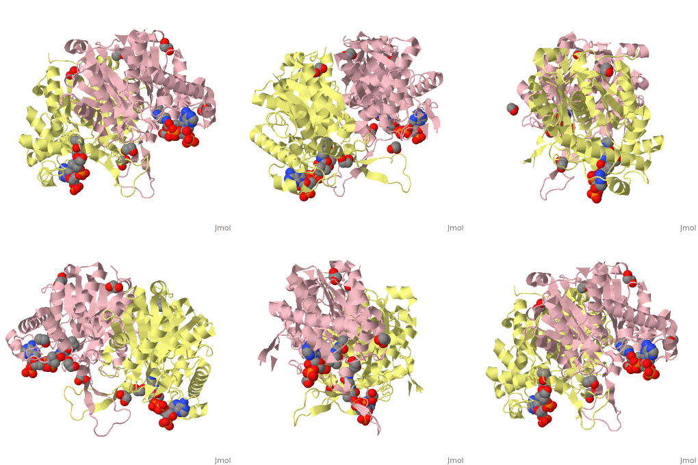
4c2j: Difference between revisions

From Proteopedia
Jump to navigation Jump to search
No edit summary
No edit summary
Line 3: Line 3:
<StructureSection load='4c2j' size='340' side='right'caption='[[4c2j]], [[Resolution|resolution]] 2.00&Aring;' scene=''>
== Structural highlights ==
<table><tr><td colspan='2'>[[4c2j]] is a 4 chain structure with sequence from [http://en.wikipedia.org/wiki/Human Human]. Full crystallographic information is available from [http://oca.weizmann.ac.il/oca-bin/ocashort?id=4C2J OCA]. For a <b>guided tour on the structure components</b> use [http://oca.weizmann.ac.il/oca-docs/fgij/fg.htm?mol=4C2J FirstGlance]. <br>
<table><tr><td colspan='2'>[[4c2j]] is a 4 chain structure with sequence from [https://en.wikipedia.org/wiki/Homo_sapiens Homo sapiens]. Full crystallographic information is available from [http://oca.weizmann.ac.il/oca-bin/ocashort?id=4C2J OCA]. For a <b>guided tour on the structure components</b> use [https://proteopedia.org/fgij/fg.htm?mol=4C2J FirstGlance]. <br>
</td></tr><tr id='ligand'><td class="sblockLbl"><b>[[Ligand|Ligands:]]</b></td><td class="sblockDat"><scene name='pdbligand=COA:COENZYME+A'>COA</scene>, <scene name='pdbligand=EDO:1,2-ETHANEDIOL'>EDO</scene></td></tr>
</td></tr><tr id='ligand'><td class="sblockLbl"><b>[[Ligand|Ligands:]]</b></td><td class="sblockDat" id="ligandDat"><scene name='pdbligand=COA:COENZYME+A'>COA</scene>, <scene name='pdbligand=CSO:S-HYDROXYCYSTEINE'>CSO</scene>, <scene name='pdbligand=EDO:1,2-ETHANEDIOL'>EDO</scene></td></tr>
<tr id='NonStdRes'><td class="sblockLbl"><b>[[Non-Standard_Residue|NonStd Res:]]</b></td><td class="sblockDat"><scene name='pdbligand=CSO:S-HYDROXYCYSTEINE'>CSO</scene></td></tr>
<tr id='resources'><td class="sblockLbl"><b>Resources:</b></td><td class="sblockDat"><span class='plainlinks'>[https://proteopedia.org/fgij/fg.htm?mol=4c2j FirstGlance], [http://oca.weizmann.ac.il/oca-bin/ocaids?id=4c2j OCA], [https://pdbe.org/4c2j PDBe], [https://www.rcsb.org/pdb/explore.do?structureId=4c2j RCSB], [https://www.ebi.ac.uk/pdbsum/4c2j PDBsum], [https://prosat.h-its.org/prosat/prosatexe?pdbcode=4c2j ProSAT]</span></td></tr>
<tr id='related'><td class="sblockLbl"><b>[[Related_structure|Related:]]</b></td><td class="sblockDat">[[4c2k|4c2k]]</td></tr>
<tr id='activity'><td class="sblockLbl"><b>Activity:</b></td><td class="sblockDat"><span class='plainlinks'>[http://en.wikipedia.org/wiki/Acetyl-CoA_C-acyltransferase Acetyl-CoA C-acyltransferase], with EC number [http://www.brenda-enzymes.info/php/result_flat.php4?ecno=2.3.1.16 2.3.1.16] </span></td></tr>
<tr id='resources'><td class="sblockLbl"><b>Resources:</b></td><td class="sblockDat"><span class='plainlinks'>[http://oca.weizmann.ac.il/oca-docs/fgij/fg.htm?mol=4c2j FirstGlance], [http://oca.weizmann.ac.il/oca-bin/ocaids?id=4c2j OCA], [http://pdbe.org/4c2j PDBe], [http://www.rcsb.org/pdb/explore.do?structureId=4c2j RCSB], [http://www.ebi.ac.uk/pdbsum/4c2j PDBsum], [http://prosat.h-its.org/prosat/prosatexe?pdbcode=4c2j ProSAT]</span></td></tr>
</table>
</table>
== Function ==
== Function ==
[[http://www.uniprot.org/uniprot/THIM_HUMAN THIM_HUMAN]] Abolishes BNIP3-mediated apoptosis and mitochondrial damage.<ref>PMID:18371312</ref
[[https://www.uniprot.org/uniprot/THIM_HUMAN THIM_HUMAN]] Abolishes BNIP3-mediated apoptosis and mitochondrial damage.<ref>PMID:18371312</ref>  
<div style="background-color:#fffaf0;">
== Publication Abstract from PubMed ==
Crystal structures of human mitochondrial 3-ketoacyl-CoA thiolase (hT1) in the apo form and in complex with CoA have been determined at 2.0 A resolution. The structures confirm the tetrameric quaternary structure of this degradative thiolase. The active site is surprisingly similar to the active site of the Zoogloea ramigera biosynthetic tetrameric thiolase (PDB entries 1dm3 and 1m1o) and different from the active site of the peroxisomal dimeric degradative thiolase (PDB entries 1afw and 2iik). A cavity analysis suggests a mode of binding for the fatty-acyl tail in a tunnel lined by the Nbeta2-Nalpha2 loop of the adjacent subunit and the Lalpha1 helix of the loop domain. Soaking of the apo hT1 crystals with octanoyl-CoA resulted in a crystal structure in complex with CoA owing to the intrinsic acyl-CoA thioesterase activity of hT1. Solution studies confirm that hT1 has low acyl-CoA thioesterase activity for fatty acyl-CoA substrates. The fastest rate is observed for the hydrolysis of butyryl-CoA. It is also shown that T1 has significant biosynthetic thiolase activity, which is predicted to be of physiological importance.
 
The crystal structure of human mitochondrial 3-ketoacyl-CoA thiolase (T1): insight into the reaction mechanism of its thiolase and thioesterase activities.,Kiema TR, Harijan RK, Strozyk M, Fukao T, Alexson SE, Wierenga RK Acta Crystallogr D Biol Crystallogr. 2014 Dec 1;70(Pt 12):3212-25. doi:, 10.1107/S1399004714023827. Epub 2014 Nov 22. PMID:25478839<ref>PMID:25478839</ref>
 
From MEDLINE&reg;/PubMed&reg;, a database of the U.S. National Library of Medicine.<br>
</div>
<div class="pdbe-citations 4c2j" style="background-color:#fffaf0;"></div>


==See Also==
==See Also==
*[[Thiolase|Thiolase]]
*[[Thiolase 3D structures|Thiolase 3D structures]]
== References ==
== References ==
<references/>
<references/>
__TOC__
__TOC__
</StructureSection>
</StructureSection>
[[Category: Acetyl-CoA C-acyltransferase]]
[[Category: Homo sapiens]]
[[Category: Human]]
[[Category: Large Structures]]
[[Category: Large Structures]]
[[Category: Harijan, R K]]
[[Category: Harijan RK]]
[[Category: Kiema, T R]]
[[Category: Kiema T-R]]
[[Category: Wierenga, R K]]
[[Category: Wierenga RK]]
[[Category: Fatty acid metabolism]]
[[Category: Mitochondrial beta-oxidation]]
[[Category: Thiolytic cleavage]]
[[Category: Transferase]]

Revision as of 20:22, 7 September 2022

Crystal structure of human mitochondrial 3-ketoacyl-CoA thiolase in complex with CoACrystal structure of human mitochondrial 3-ketoacyl-CoA thiolase in complex with CoA

Structural highlights

4c2j is a 4 chain structure with sequence from Homo sapiens. Full crystallographic information is available from OCA. For a guided tour on the structure components use FirstGlance.
Ligands:, ,
Resources:FirstGlance, OCA, PDBe, RCSB, PDBsum, ProSAT

Function

[THIM_HUMAN] Abolishes BNIP3-mediated apoptosis and mitochondrial damage.[1]

See Also

References

  1. Cao W, Liu N, Tang S, Bao L, Shen L, Yuan H, Zhao X, Lu H. Acetyl-Coenzyme A acyltransferase 2 attenuates the apoptotic effects of BNIP3 in two human cell lines. Biochim Biophys Acta. 2008 Jun;1780(6):873-80. doi: 10.1016/j.bbagen.2008.02.007., Epub 2008 Mar 6. PMID:18371312 doi:http://dx.doi.org/10.1016/j.bbagen.2008.02.007

4c2j, resolution 2.00Å

Drag the structure with the mouse to rotate

Proteopedia Page Contributors and Editors (what is this?)Proteopedia Page Contributors and Editors (what is this?)

OCA