2fom: Difference between revisions
No edit summary |
No edit summary |
||
| Line 3: | Line 3: | ||
<StructureSection load='2fom' size='340' side='right'caption='[[2fom]], [[Resolution|resolution]] 1.50Å' scene=''> | <StructureSection load='2fom' size='340' side='right'caption='[[2fom]], [[Resolution|resolution]] 1.50Å' scene=''> | ||
== Structural highlights == | == Structural highlights == | ||
<table><tr><td colspan='2'>[[2fom]] is a 2 chain structure with sequence from [ | <table><tr><td colspan='2'>[[2fom]] is a 2 chain structure with sequence from [https://en.wikipedia.org/wiki/Dengue_virus_2 Dengue virus 2]. Full crystallographic information is available from [http://oca.weizmann.ac.il/oca-bin/ocashort?id=2FOM OCA]. For a <b>guided tour on the structure components</b> use [https://proteopedia.org/fgij/fg.htm?mol=2FOM FirstGlance]. <br> | ||
</td></tr><tr id='ligand'><td class="sblockLbl"><b>[[Ligand|Ligands:]]</b></td><td class="sblockDat"><scene name='pdbligand=CL:CHLORIDE+ION'>CL</scene>, <scene name='pdbligand=GOL:GLYCEROL'>GOL</scene></td></tr> | </td></tr><tr id='ligand'><td class="sblockLbl"><b>[[Ligand|Ligands:]]</b></td><td class="sblockDat" id="ligandDat"><scene name='pdbligand=CL:CHLORIDE+ION'>CL</scene>, <scene name='pdbligand=GOL:GLYCEROL'>GOL</scene></td></tr> | ||
<tr id='activity'><td class="sblockLbl"><b>Activity:</b></td><td class="sblockDat"><span class='plainlinks'>[ | <tr id='activity'><td class="sblockLbl"><b>Activity:</b></td><td class="sblockDat"><span class='plainlinks'>[https://en.wikipedia.org/wiki/Flavivirin Flavivirin], with EC number [https://www.brenda-enzymes.info/php/result_flat.php4?ecno=3.4.21.91 3.4.21.91] </span></td></tr> | ||
<tr id='resources'><td class="sblockLbl"><b>Resources:</b></td><td class="sblockDat"><span class='plainlinks'>[ | <tr id='resources'><td class="sblockLbl"><b>Resources:</b></td><td class="sblockDat"><span class='plainlinks'>[https://proteopedia.org/fgij/fg.htm?mol=2fom FirstGlance], [http://oca.weizmann.ac.il/oca-bin/ocaids?id=2fom OCA], [https://pdbe.org/2fom PDBe], [https://www.rcsb.org/pdb/explore.do?structureId=2fom RCSB], [https://www.ebi.ac.uk/pdbsum/2fom PDBsum], [https://prosat.h-its.org/prosat/prosatexe?pdbcode=2fom ProSAT]</span></td></tr> | ||
</table> | </table> | ||
== Function == | == Function == | ||
[[ | [[https://www.uniprot.org/uniprot/Q91H74_9FLAV Q91H74_9FLAV]] Envelope protein E binding to host cell surface receptor is followed by virus internalization through clathrin-mediated endocytosis. Envelope protein E is subsequently involved in membrane fusion between virion and host late endosomes. Synthesized as a homodimer with prM which acts as a chaperone for envelope protein E. After cleavage of prM, envelope protein E dissociate from small envelope protein M and homodimerizes (By similarity).[SAAS:SAAS026470_004_099774] | ||
== Evolutionary Conservation == | == Evolutionary Conservation == | ||
[[Image:Consurf_key_small.gif|200px|right]] | [[Image:Consurf_key_small.gif|200px|right]] | ||
| Line 29: | Line 29: | ||
</div> | </div> | ||
<div class="pdbe-citations 2fom" style="background-color:#fffaf0;"></div> | <div class="pdbe-citations 2fom" style="background-color:#fffaf0;"></div> | ||
== References == | == References == | ||
<references/> | <references/> | ||
Revision as of 23:39, 20 October 2021
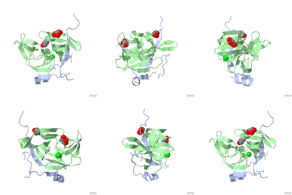
Dengue Virus NS2B/NS3 ProteaseDengue Virus NS2B/NS3 Protease
Structural highlights
Function[Q91H74_9FLAV] Envelope protein E binding to host cell surface receptor is followed by virus internalization through clathrin-mediated endocytosis. Envelope protein E is subsequently involved in membrane fusion between virion and host late endosomes. Synthesized as a homodimer with prM which acts as a chaperone for envelope protein E. After cleavage of prM, envelope protein E dissociate from small envelope protein M and homodimerizes (By similarity).[SAAS:SAAS026470_004_099774] Evolutionary Conservation Check, as determined by ConSurfDB. You may read the explanation of the method and the full data available from ConSurf. Publication Abstract from PubMedThe replication of flaviviruses requires the correct processing of their polyprotein by the viral NS3 protease (NS3pro). Essential for the activation of NS3pro is a 47-residue region of NS2B. Here we report the crystal structures of a dengue NS2B-NS3pro complex and a West Nile virus NS2B-NS3pro complex with a substrate-based inhibitor. These structures identify key residues for NS3pro substrate recognition and clarify the mechanism of NS3pro activation. Structural basis for the activation of flaviviral NS3 proteases from dengue and West Nile virus.,Erbel P, Schiering N, D'Arcy A, Renatus M, Kroemer M, Lim SP, Yin Z, Keller TH, Vasudevan SG, Hommel U Nat Struct Mol Biol. 2006 Apr;13(4):372-3. Epub 2006 Mar 12. PMID:16532006[1] From MEDLINE®/PubMed®, a database of the U.S. National Library of Medicine. References
|
| ||||||||||||||||||
